Altered sensation after lower wisdom-tooth surgery is an important consent topic, but lasting nerve injury is uncommon. Most postoperative nerve disturbances are temporary and improve over time. Rarely, injury to a sensory nerve can leave persistent numbness, tingling, burning pain, or altered taste. The risk for one person depends heavily on the tooth’s position and its relationship to nearby nerves.

Which nerves can be affected?
The nerves most often discussed with lower third-molar surgery are the inferior alveolar nerve (IAN) and the lingual nerve (LN). Both carry sensory information; routine lower wisdom-tooth extraction is not expected to paralyze the lip, tongue, or jaw. Describing these complications as typical muscle weakness or paralysis is misleading.
Symptoms may be described as numbness, pins and needles, burning, hypersensitivity, reduced sensation, or an unusual response to touch or temperature. Lingual nerve disturbance can also affect taste. A person may find eating or speaking uncomfortable when sensation is altered, but movement problems should not be presented as the expected effect of an IAN or LN sensory injury.
For background on the procedure itself, see our guides to when wisdom teeth emerge and wisdom-tooth pain. Neither a symptom article nor a photograph can estimate an individual’s nerve risk; the surgeon needs to review the examination and imaging.
How common is persistent nerve injury?
Temporary sensory changes are more common than permanent ones. AAOMS describes numbness that lasts beyond the short anesthetic period as uncommon and usually temporary, although permanent change can occur rarely. Research estimates vary because studies include different tooth positions, surgical approaches, operators, follow-up periods, and definitions of “permanent.”
One systematic review covering 44,171 impacted mandibular third-molar extractions reported an approximately 1.20% rate of transient IAN deficit and 0.28% rate of permanent IAN deficit. A separate 2023 review of lingual nerve outcomes reported study-level mean permanent-injury rates that differed across three surgical approaches, from about 0.07% to 0.28%. These figures describe groups in published studies—not a personal risk calculator, and not a guarantee for an individual procedure.
Risk estimates for lower wisdom teeth should not be applied to every tooth extraction or to upper wisdom teeth. If a study reports that a symptom was still present at a particular follow-up point, that does not by itself predict that the symptom will remain forever. The surgeon can explain how the published ranges relate, or do not relate, to the patient’s anatomy and the planned procedure.
What changes a person’s risk?
The relationship between a lower wisdom tooth’s roots and the mandibular canal is one of the most important anatomic considerations for IAN injury. The depth and angle of impaction, the amount of bone covering the tooth, the surgical field, and technique can also matter. Age may be associated with risk in some datasets, but it does not replace evaluation of the actual tooth and nerve position.
Experience and appropriate training are relevant to planning and technique. At the same time, a complication does not prove that a surgeon was unskilled: a nerve can sit very close to the tooth, and even carefully planned surgery may carry a risk. A useful consultation focuses on the images, the anticipated difficulty, who will perform the procedure, and how the team will respond if sensory changes occur.
Other health factors may affect surgical planning or healing, so give the oral surgeon an accurate medical history and medication list. These issues do not all directly cause nerve injury. They help the care team choose a plan that fits the whole patient rather than relying on a generic warning.
How imaging helps plan lower wisdom-tooth surgery
A panoramic radiograph is commonly used to assess impacted wisdom teeth and their relationship to nearby structures. If the roots appear close to the inferior alveolar canal and three-dimensional detail could change risk assessment or management, the surgeon may consider cone-beam computed tomography (CBCT). CBCT is not automatically needed for every patient, and additional imaging should answer a clinical question rather than be ordered as a routine precaution without a reason.
Imaging can help clarify the canal’s position and guide a conversation about full removal, monitoring, or an alternative such as coronectomy. It cannot make surgery risk-free or predict with certainty whether a nerve will recover. If you are unsure why a scan is recommended, ask what finding on the existing image prompted it and how the result might affect the treatment plan.
For additional context, see our pages on wisdom-tooth development and wisdom-tooth anatomy. These explain general development and anatomy; your own radiographs remain the relevant source for individual surgical planning.
When coronectomy may be discussed
For selected lower wisdom teeth whose roots are very close to the IAN, an oral surgeon may discuss coronectomy. In this procedure the crown is removed while suitable roots are intentionally retained, with the aim of reducing the need to disturb the nerve-side root region. It is not appropriate for every tooth. Decay, infection, root mobility, and other clinical factors may make it unsuitable.
Systematic reviews report low IAN-injury rates in coronectomy series, but the evidence includes different techniques and study designs. Retained roots can migrate, become exposed, or occasionally require later treatment. The patient and surgeon should weigh those possibilities against the risk of complete extraction for that particular tooth. AAOMS offers a patient overview of when coronectomy may be considered.
Monitoring may be another option for some wisdom teeth, depending on symptoms, pathology, and the patient’s circumstances. Removal is not automatically the right choice for every asymptomatic tooth. A discussion of the reason for treatment and the alternatives is part of informed consent.
When to report numbness, tingling, or taste changes
Local anesthetic temporarily numbs part of the mouth. Sensation that is absent during the expected anesthetic period is not enough to diagnose permanent nerve damage. If the lower lip, chin, or tongue still feels clearly abnormal after the anesthetic should have worn off, contact the oral surgeon promptly. Describe exactly where sensation changed, when it began, and whether it is improving, stable, or worsening.
Do not wait months to mention a dense numb area, painful burning, severe touch sensitivity, or a deficit that is not improving. Early documentation and follow-up allow the clinician to compare the sensory territory and recovery over time. This does not mean that early numbness is permanent; sensory recovery may continue for weeks or months.
- Where the sensation changed: lower lip, chin, tongue, or another area.
- What it feels like: numb, tingling, burning, painful, unusually sensitive, or altered taste.
- When it started and whether it is improving, staying the same, or worsening.
For general recovery expectations after extraction, read our guide to wisdom-tooth signs and symptoms and the overview of potential wisdom-tooth complications. If you notice a sensory deficit after your own procedure, contact the treating surgeon rather than relying on a general article.
How a clinician evaluates a sensory change
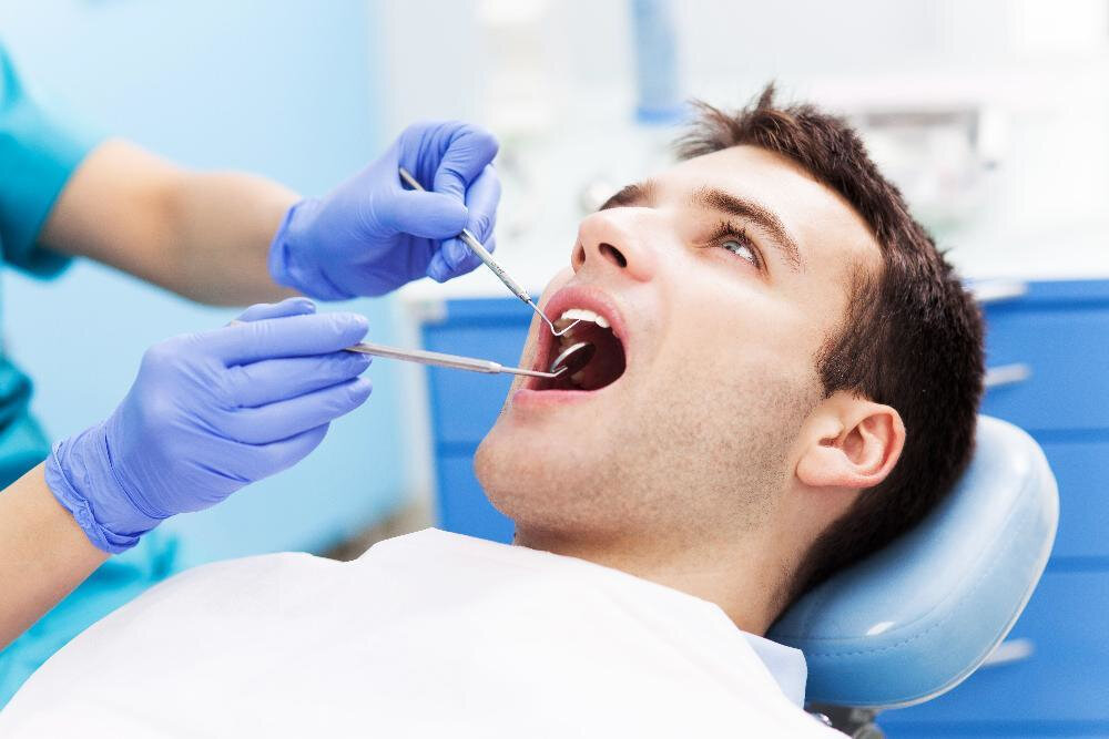
Assessment usually starts with a detailed history and a clinical neurosensory examination. The clinician may compare the left and right sides, map the area that feels different, and check responses to light touch, direction, pinprick, or temperature as appropriate. This helps distinguish an IAN-pattern change from a lingual-nerve pattern and creates a baseline for follow-up.
There is no single test that is the default for every dental nerve concern. Additional imaging or other tests depend on the suspected nerve injury and whether the result would change care. Limb-style electromyography or nerve-conduction studies are not a routine first-line workup for every post-extraction sensory symptom.
Bring the operative instructions and explain whether the change is numbness, tingling, burning, pain, altered taste, or reduced sensitivity. A clear description is more useful than labeling the problem “permanent” in the first days after surgery.
Management and specialist referral
Early care may include observation with repeat sensory testing and follow-up appointments. If painful post-traumatic nerve symptoms are present, a clinician can assess whether targeted neuropathic-pain treatment is appropriate. Ordinary pain medicine may help expected surgical soreness, but it does not repair a nerve. Do not start prescription medicines, supplements, or procedures as self-treatment.
Persistent, significant IAN or lingual-nerve deficits may warrant assessment by an oral and maxillofacial surgeon or a clinician with microsurgical nerve expertise. Referral timing and treatment candidacy depend on which nerve is affected, the severity and type of deficit, pain or taste symptoms, and whether sensation is recovering. A specialist may discuss continued observation, nerve repair, grafting, or other options in selected cases.
A 2026 systematic review found favorable functional sensory recovery in many reported cases after intervention, but the evidence is heterogeneous and drawn from selected patients who received treatment. Its pooled outcomes cannot promise a result for an individual or determine who should have surgery. Regenerative treatments such as stem cells remain research topics, not established standard care for wisdom-tooth nerve injury. Acupuncture or chiropractic care should not be presented as repairing a surgically injured trigeminal nerve.
Persistent numbness or painful altered sensation can affect eating, speaking comfortably, sleep, and quality of life. If symptoms are distressing, ask the treating team about pain management and psychological support as part of care. Support addresses the impact of symptoms; it does not imply that the symptoms are imagined.
Questions to ask before surgery
Informed consent is a two-way discussion. Ask the surgeon to explain the reason for the procedure, the relevant anatomy, the likely benefits, the alternatives, and the specific risks suggested by your imaging. Useful questions include:
- Are the roots close to the inferior alveolar canal or lingual nerve on my images?
- Would a CBCT scan change the risk assessment or treatment plan in my case?
- Is full removal, coronectomy, or monitoring reasonable for this tooth, and why?
- Who will perform the surgery, and how will sensory changes be checked afterward?
- Whom should I contact if numbness, tingling, burning pain, or taste changes remain after the anesthetic wears off?
Choosing a trained, appropriately experienced clinician and discussing the plan can help you understand the procedure. Ask for the individualized explanation rather than a generic promise of a particular “success rate.” A clinician should be able to explain uncertainty and answer what follow-up would look like.
Common questions
Takeaway
Permanent sensory nerve injury after lower wisdom-tooth removal is rare; temporary altered sensation is more common and often improves. Risk depends heavily on anatomy, especially how the roots relate to the inferior alveolar canal, as well as tooth position and the surgical plan. Imaging and, for selected high-risk teeth, coronectomy may inform risk reduction, but neither guarantees an outcome. Report persistent lower-lip, chin, or tongue sensory changes promptly so the oral surgeon can document and follow recovery, with specialist referral when symptoms are significant or improvement is poor.