What cellular senescence is
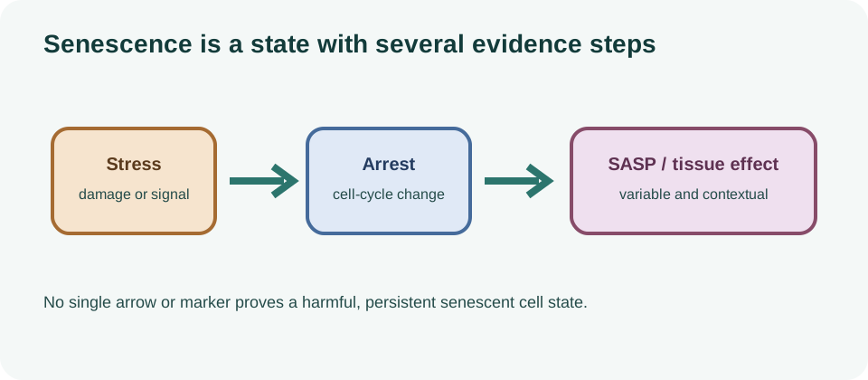
Cellular senescence describes a state in which a cell has durably reduced or stopped proliferation while remaining metabolically active. The state can involve chromatin remodeling, altered metabolism, lysosomal changes, DNA-damage signaling, resistance to apoptosis and a senescence-associated secretory phenotype (SASP). No single feature is present in every senescent cell, and no single marker is specific enough to define the state across tissues.
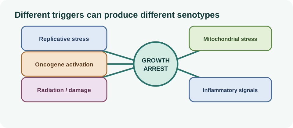
Senescence is therefore better understood as a functional phenotype than as a permanent cell identity. A cell can enter the state after replicative exhaustion, DNA damage, oncogene activation, mitochondrial stress, irradiation, chemotherapy or other perturbations. Its molecular profile depends on the trigger, cell type, tissue environment, time since induction and ability of the immune system to remove it.
That distinction replaces the popular “zombie cell” metaphor with a more useful question: what state is present, in which cells, for how long, with what secretory program, and with what effect on tissue function?
Growth arrest is necessary but not sufficient
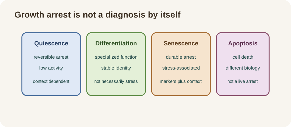
Senescent cells commonly show reduced proliferation, but growth arrest also occurs in quiescence, terminal differentiation, contact inhibition, temporary stress responses and some forms of dormancy. A non-dividing cell is not automatically senescent. Conversely, senescence-like cells can be difficult to classify if the arrest is incomplete or the sample contains several cell states.
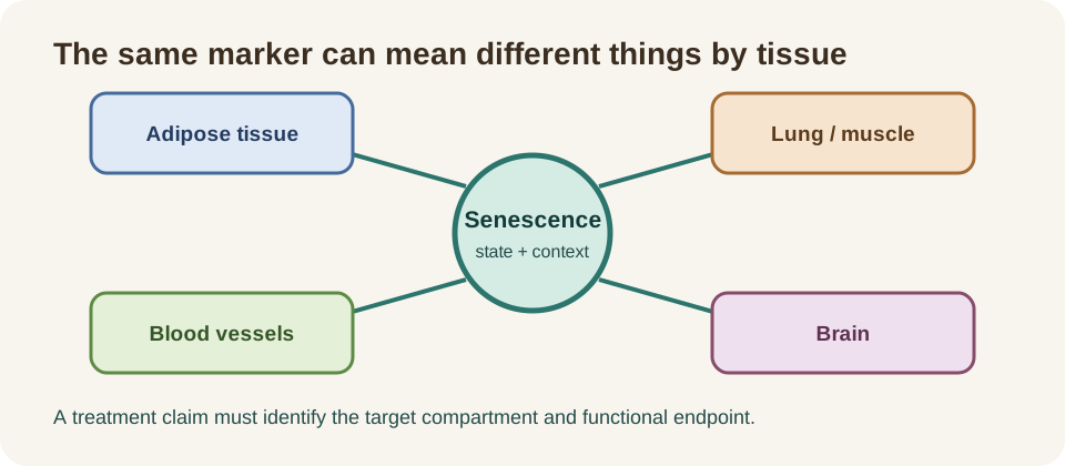
Researchers may combine markers such as p16INK4a, p21CIP1, persistent DNA-damage foci, altered lysosomal activity, enlarged morphology, anti-apoptotic signaling and selected SASP factors. The combination must be validated in the relevant tissue. A marker that works in cultured fibroblasts may perform differently in adipose tissue, lung, blood or brain.
| State | Typical feature | Why it can be confused with senescence |
|---|---|---|
| Quiescence | reversible low proliferation during limited growth signals. | the cell is not dividing, but may re-enter the cycle. |
| Terminal differentiation | specialized mature function with little division. | durable arrest can resemble a senescent phenotype. |
| Senescence | stress-associated durable arrest plus context-dependent remodeling. | the phenotype is heterogeneous and lacks one universal marker. |
| Apoptosis | regulated cell death. | both can follow severe stress, but their tissue effects differ. |
Replicative, DNA-damage, oncogene and stress-induced senescence
Replicative senescence can emerge after repeated division and telomere-associated stress. DNA-damage-induced senescence follows genotoxic injury or persistent replication stress. Oncogene-induced senescence is a protective response that can restrain cells with abnormal proliferative signaling. Other forms arise from mitochondrial dysfunction, oxidative stress, nutrient imbalance, inflammation, irradiation or drug exposure.
These labels describe experimental routes, not four perfectly separate diseases. Different triggers can converge on p53–p21, p16–RB, chromatin, mitochondrial and inflammatory pathways, while retaining distinct transcriptional and secretory programs. A drug-induced senescence model may therefore resemble, but not reproduce, the state that accumulates during aging in a human tissue.
Timing also matters. A transient state may resolve, be cleared by immune cells, or be remodeled into a persistent state. A tissue sample taken at one time point cannot always distinguish those trajectories.
Transient beneficial senescence versus chronic persistence
Senescence is not inherently harmful. During development, transient senescence can help shape tissues. During wound healing, a temporary senescent program may coordinate remodeling and recruit immune cells. In tumor suppression, durable arrest can prevent a damaged or oncogene-activated cell from continuing to divide.
The problem is persistence, burden and context. If senescent cells are not cleared or if their secretory program remains active, they can alter neighboring cells, extracellular matrix, stem-cell niches and immune signaling. Yet removing every cell with a senescence marker could interfere with repair, tumor suppression or normal tissue homeostasis.
The appropriate therapeutic goal is therefore not “eliminate senescence everywhere.” It is to identify harmful persistent states, preserve useful transient functions and intervene in a tissue- and disease-specific way.
The SASP is not one fixed molecular signature
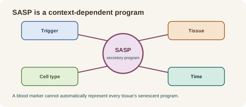
The SASP refers to a collection of secreted cytokines, chemokines, growth factors, proteases, extracellular vesicles and other molecules associated with some senescent states. It can influence neighboring cells and immune recruitment. The composition changes with the inducing stress, cell lineage, culture conditions, time and inflammatory environment.
Some SASP factors are also produced by macrophages, injured cells, infections and ordinary inflammatory responses. A high circulating cytokine therefore does not identify a senescent source. Conversely, a senescent cell may have a modest or unusual secretome that is missed by a standard panel.
Recent human biomarker reviews highlight circulating candidates such as GDF15 and Activin A as potentially useful across health domains, but usefulness is not specificity. A biomarker panel may predict risk or correlate with disease without proving how many senescent cells are present or whether senolytic treatment would help.
| Observation | Possible meaning | What it does not prove |
|---|---|---|
| Circulating cytokine rises | inflammation, tissue stress or a senescence-associated signal. | that senescent cells are the only source. |
| Panel changes after treatment | altered secretion or systemic state. | selective clearance of harmful cells. |
| Low SASP signal | low secretion, timing or assay limitation. | absence of senescence in every tissue. |
Senotype heterogeneity across tissues and cell types
“Senotype” is a useful shorthand for the molecular and functional profile of a senescent cell in its context. Endothelial, epithelial, stromal, immune, adipocyte, muscle and neural cells can enter different states. Even within one tissue, cells may differ in trigger, burden, SASP, immune visibility and response to a drug.
The 2026 Cell framework for charting human cellular senescence emphasizes cell-type-resolved and tissue-aware approaches. This matters because a blood signature can mix signals from multiple organs, while a biopsy samples only a small region. A whole-body senescence score can be attractive to consumers but should not be treated as a validated diagnosis without rigorous longitudinal testing.
Heterogeneity also explains why a senolytic that works in a mouse model or one human disease may not work in another. Drug sensitivity, uptake, anti-apoptotic dependence and neighboring-cell effects are not universal properties of all senescent cells.
Immune surveillance and clearance
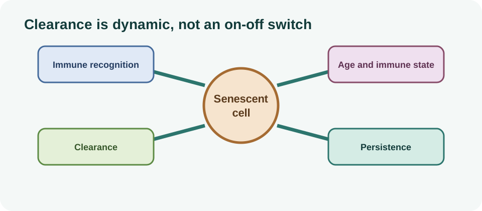
Natural killer cells, macrophages and other immune mechanisms can recognize and remove some senescent cells. Aging, chronic inflammation, tissue barriers, altered antigen presentation and senescent-cell resistance to apoptosis may reduce effective clearance. But immune recruitment is not always beneficial: an inflammatory response can damage tissue, and some senescent programs may help organize repair.
Clearance is therefore a dynamic balance between formation, persistence, immune access and removal. A rise in a senescence marker after injury may reflect active repair and immune surveillance rather than an irreversible pathological burden. A fall after treatment may reflect altered secretion or sampling rather than selective killing.
Inflammaging, fibrosis and secondary senescence
Persistent senescent cells can release signals that activate fibroblasts, alter extracellular matrix turnover, recruit immune cells and change stem-cell niches. These pathways may contribute to chronic low-grade inflammation, fibrosis, impaired regeneration and tissue dysfunction. The related inflammaging page explains why an inflammatory marker is a broad signal rather than a senescence diagnosis.
Senescent-cell secretions can also induce senescence-like changes in neighboring cells, a process often called paracrine or secondary senescence. The direction and importance of that spread depend on the factor, dose, tissue and duration. It should not be inferred from a cell-culture observation that a whole human organ is undergoing the same process.
These effects intersect with proteostasis, mitochondrial quality, extracellular matrix biology and stem-cell exhaustion. A single hallmark rarely explains the clinical phenotype by itself.
Senescence in adipose tissue, vessels, lung, muscle and brain
In adipose tissue, senescent stromal or precursor cells may influence adipokines, inflammation and regenerative capacity. In the vasculature, endothelial and smooth-muscle states may interact with oxidative stress, plaque biology and repair. In the lung, epithelial and fibroblast senescence has been studied in fibrosis and injury. These examples do not imply the same senotype or intervention across organs.
Muscle aging involves satellite cells, fibro-adipogenic progenitors, immune cells, neuromuscular interactions and systemic metabolism. A senescence marker in a muscle sample cannot be equated with sarcopenia, and removing a cell population without understanding its repair role could be harmful.
In the brain, glial and vascular states may influence neuroinflammation and neuronal support, but the blood–brain barrier, cellular diversity and sampling limits make translation difficult. The brain-aging page provides context for why a peripheral biomarker is not automatically a brain measurement.
How senescence is measured in humans
Human studies use combinations of tissue staining, immunohistochemistry, transcriptomics, proteomics, circulating factors, cell-surface markers, single-cell sequencing, spatial profiling and functional assays. Each method answers a different question. A plasma assay is scalable, a biopsy provides local information, and single-cell or spatial methods can reveal which cells carry a signal.
Strong studies report sample handling, cell composition, assay validation, thresholds, batch effects, missingness and replication. They also connect a molecular signature to a clinical or functional outcome. A cross-sectional association between a marker and age is not enough to establish causation, persistence or treatment response.
| Marker or method | Potential use | Important limitation |
|---|---|---|
| p16 or p21 | evidence of cell-cycle and stress-pathway activity. | not universal or specific as a stand-alone marker. |
| SA-β-gal | lysosomal activity in appropriate experimental settings. | context- and assay-dependent; not a whole-body diagnosis. |
| Circulating SASP factors | accessible signals associated with tissue and disease states. | many are produced by non-senescent cells and inflammation. |
| Single-cell/spatial profile | cell-type and location-resolved state classification. | cost, sampling, analysis and longitudinal validation remain limiting. |
Why p16, p21 and SA-β-gal are not universal stand-alone markers
p16 and p21 are central regulators of cell-cycle arrest, but their expression can change in non-senescent states and may be absent from some senescent cells. SA-β-gal reflects lysosomal activity and can be useful in defined assays, but its interpretation changes with cell type, pH, culture conditions and tissue processing. SASP factors have the same specificity problem in blood.
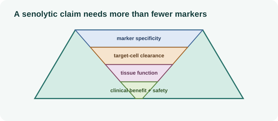
Using a panel improves confidence only if the panel is validated. Counting more markers does not automatically solve the problem if they are biologically redundant, measured in the wrong compartment or selected after looking at the result. Functional evidence—durable arrest, altered survival, secretory behavior, tissue effect and response to a defined intervention—adds the missing context.
| Approach | Added information | Remaining requirement |
|---|---|---|
| Single-cell | which cell types carry a candidate state. | functional and longitudinal validation. |
| Spatial | where the state sits and which neighbors it contacts. | representative sampling and outcome linkage. |
| Multi-omic | convergent molecular layers beyond one marker. | independent cohorts and interpretable classifiers. |
Single-cell, spatial and multi-omic approaches
Single-cell RNA sequencing can separate cell types and identify state-specific gene programs. Spatial transcriptomics and imaging retain the tissue neighborhood, which is important for paracrine effects, immune access and fibrosis. Proteomics, epigenomics and metabolomics add layers that transcript counts cannot capture alone.
These tools are changing the field from “find the senescence gene” to “classify a state in a cell, location and time.” The 2026 SENESCENCE2030 roadmap calls for functional classifications, standardized clinically actionable biomarkers, precision translation and stronger regulatory frameworks. That is a research agenda, not proof that consumer senescence tests or universal senolytic schedules are ready.
Multi-omic complexity also needs restraint. A machine-learning signature can predict age or disease while remaining difficult to interpret, vulnerable to cohort shift and unvalidated as a treatment-selection tool. Independent cohorts and prospective intervention studies are essential.
Where senolytics and senomorphics fit
Senolytics aim to selectively eliminate vulnerable senescent cells. Senomorphics, sometimes called senostatics, aim to modify harmful secretory or survival behavior without necessarily killing the cell. Immune-mediated clearance and tissue-specific delivery are additional strategies. The choice depends on the senotype, disease, tissue and safety margin.
Early clinical studies are encouraging in selected conditions, but they do not establish a general anti-aging treatment. A candidate can improve a symptom or function without proving that senescent-cell burden was the causal mediator. Conversely, a biomarker change without better function is not enough.
The senolytics human-evidence page tracks this distinction. The field should preserve beneficial senescence, avoid interference with wound repair and tumor suppression, and monitor infection, tissue injury, hematologic effects and other long-term risks.
Evidence verdict and translational priorities
Cellular senescence is a real and important biology of aging, but it is not a single uniform cell type and it is not always pathological. Persistent states can contribute to inflammation, fibrosis, stem-cell dysfunction and tissue decline, while transient states can support development, repair and tumor suppression. The central human limitation remains classification: no universal marker identifies every relevant senescent cell or tells us whether it should be removed.
Progress requires validated cell-type and tissue-resolved signatures, longitudinal sampling, functional assays, independent cohorts and clinical trials with meaningful outcomes. Future interventions should be precision strategies aimed at harmful persistent states, not blanket clearance. Findings should remain useful even when one marker or model is later revised.
- define senescence with multiple validated features and functional context;
- separate quiescence, differentiation, injury and senescence;
- identify the cell type, tissue, trigger and duration;
- connect molecular classification to patient-important function;
- preserve null, contradictory and safety findings in the evidence record.