The circadian timing system: one coordinator and many local clocks
“Circadian” means occurring on an approximately daily cycle. The word does not mean that every measured rhythm lasts exactly 24 hours or that every process follows the same schedule. An internal timing system generates recurring patterns, while environmental and behavioral signals synchronize those patterns with the day-night cycle.
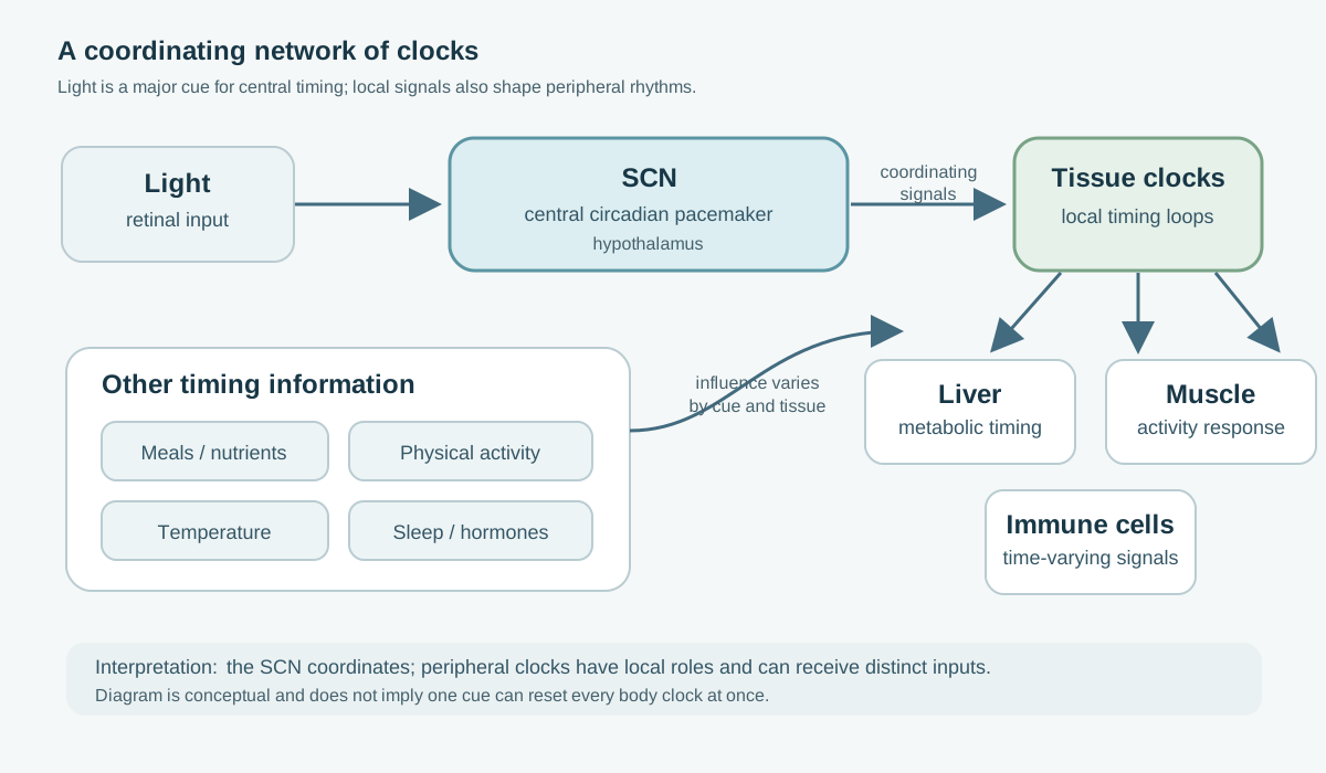
The suprachiasmatic nucleus, or SCN, is a small group of neurons in the hypothalamus. Specialized retinal pathways convey information about ambient light to the SCN. That information helps the brain align internal timing with the outside light-dark cycle. The SCN sends neural, hormonal and behavioral timing signals that help coordinate rhythms throughout the body. It is central to the system, but it does not micromanage every cell or determine all daily physiology on its own.
Cells in many tissues also contain molecular clock machinery. These peripheral clocks help time functions such as liver metabolism, muscle activity, immune signaling and hormone responsiveness. Light is a dominant cue for the brain’s central clock. Meal timing, physical activity, temperature, sleep-wake behavior and social routines can also provide timing information, with effects that depend on tissue and context. These cues are sometimes called zeitgebers, or time-givers. They are not interchangeable: a meal can shift some peripheral signals without necessarily shifting the SCN by the same amount.
This distinction matters for interpreting claims about “resetting the body clock.” There is no single clock hand that can be moved with one universal intervention. A change in sleep timing, melatonin onset, activity regularity or a gene-expression rhythm may describe different parts of a coupled timing system.
How molecular clocks produce recurring patterns
At the cellular level, circadian timing depends on interacting gene and protein feedback loops. In a simplified description, CLOCK and BMAL1 proteins activate the expression of genes including PER and CRY. As PER and CRY proteins accumulate, they feed back to inhibit CLOCK-BMAL1 activity. Protein breakdown and additional regulatory loops then allow the cycle to begin again. The timing emerges from a network of transcription, translation, chemical modification and protein turnover.
This molecular loop is not a tiny mechanical timer with one perfectly stable period. Its timing is affected by cell state, temperature, nutrients, hormones and signals from neighboring cells. In a tissue, many cells can become more or less synchronized with one another. Across the body, local clocks can maintain different phases because organs have different jobs and receive different cues.
Daily rhythms are also not proof that the molecular clock caused a particular health outcome. A blood measurement may vary over the day because of a circadian process, a meal, posture, sleep, activity or their combination. Experimental protocols such as constant routine or forced desynchrony attempt to separate endogenous circadian effects from immediate behavior, but they are demanding and involve small samples. A field study can be more representative of ordinary life while being less able to identify the source of a rhythm.
| Property | What it describes | What it cannot establish alone |
|---|---|---|
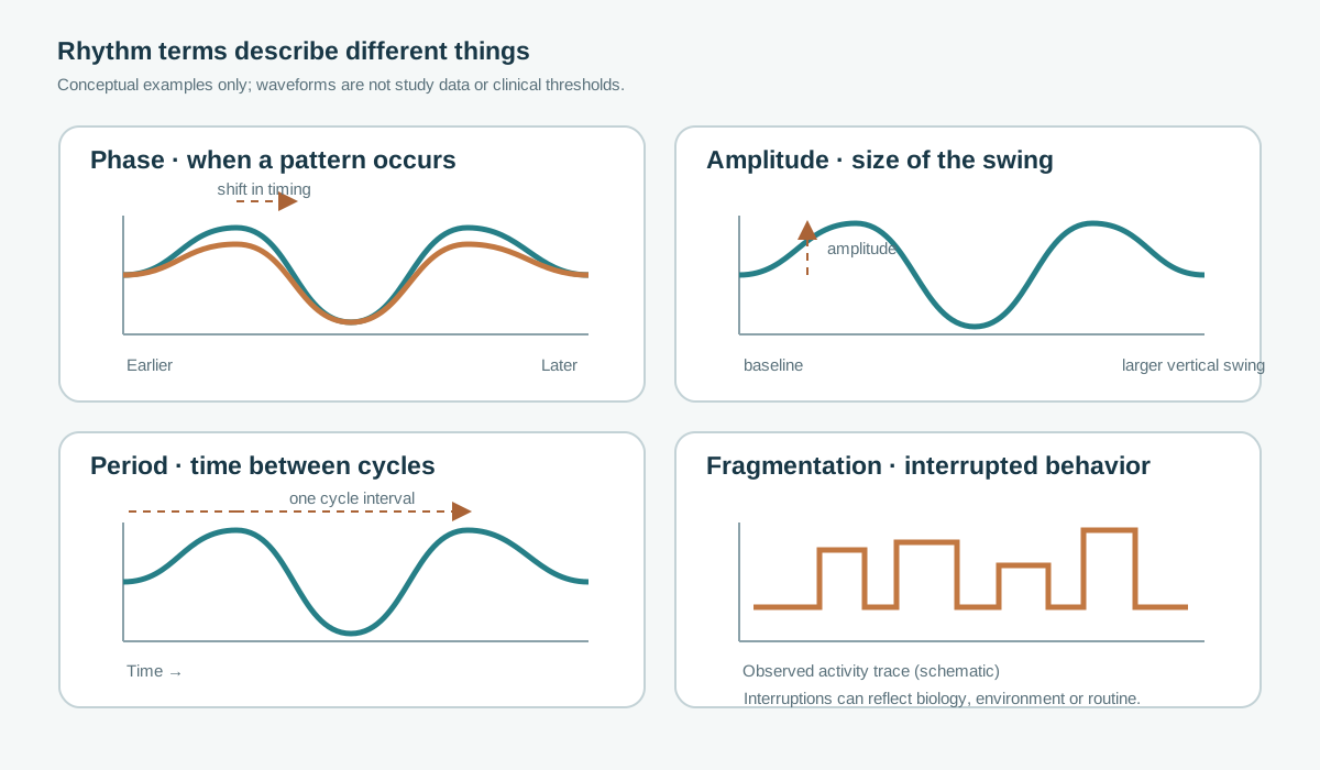
| Phase | Where a recurring pattern falls in time, such as the onset of melatonin under dim light | Whether the rhythm is strong, healthy or correctly aligned for a specific person |
| Amplitude | The size of a rhythm’s rise and fall under a stated measurement method | That all tissues share the same amplitude or that a larger value is always better |
| Period | The time between cycles generated by an oscillator | That observed behavior follows the endogenous period without environmental influence |
| Regularity / fragmentation | How consistently an observed behavior or physiological signal recurs, and how often it is interrupted | A direct measurement of the SCN or of molecular clocks in every tissue |
What may change with age: phase, amplitude, fragmentation and synchrony
Some human studies find that sleep and circadian phase occur earlier on average in older than younger groups. Other studies report lower amplitude in selected rhythms, more variable day-to-day activity, or weaker alignment between signals. These findings are not a universal aging signature. Results differ across melatonin, temperature, cortisol, activity and metabolic measures, and can be affected by health status, medications, light exposure, sex, living environment and study design.
A scoping review of 42 eligible studies on body-fluid rhythms in older people found that many studies reported flattening for some measured parameters, but the literature was heterogeneous and included contradictory results; several parameters appeared in only one study. This is a useful description of the field’s limits: “aging weakens every circadian rhythm” is too broad. A 2023 controlled lipidomics analysis of 24 healthy participants—12 younger adults and 12 middle-aged participants, not a representative sample of the oldest old—found that some lipid rhythms persisted, while the older group showed lower average amplitude and earlier acrophase for the rhythmic lipids studied. Its narrow age groups and small sample make it a mechanistic observation, not a population-wide estimate.
Fragmentation adds another layer. A person can have a relatively stable internal phase while sleep or activity is repeatedly interrupted by caregiving, pain, nocturia, shift work or an irregular environment. Conversely, a regular bedtime does not prove that the internal circadian phase is aligned with that schedule. The observed pattern is produced by both the internal system and a person’s circumstances.
Fragmentation is not the same as phase change
Synchrony can refer to relationships among cells in one tissue, among tissues, or between internal rhythms and external demands. It is harder to measure than a single blood or wearable signal. Human evidence often uses proxies such as activity rhythms or the timing of one hormone. Those proxies are informative for their specific question, but they do not directly measure coordination among every organ clock.
Sleep, circadian phase, chronotype and regularity are related but distinct
Sleep is shaped by at least two interacting processes: circadian timing and homeostatic sleep pressure, which builds during wakefulness and dissipates during sleep. The circadian system helps create daily windows when sleep is more or less likely. Homeostatic pressure helps explain why a long period awake makes sleep more likely even when the circadian phase is not at its usual sleep-promoting time.
Sleep duration is the total amount of time asleep. Sleep timing is when sleep occurs on the clock. Circadian phase is estimated from physiological markers such as dim-light melatonin onset (DLMO), not simply from bedtime. Chronotype is a behavioral tendency toward earlier or later timing, usually assessed by questionnaires or sleep timing. Activity regularity describes patterns in movement across days. These measures can correlate without being interchangeable.
For example, an older adult may go to bed early because of preference, a medication, reduced evening activity, caregiving demands or an earlier circadian phase. A wrist actigraph can estimate rest-activity patterns but cannot diagnose which explanation is correct. A DLMO assessment can estimate internal phase under controlled dim light but does not tell the whole story about tissue clocks or sleep quality.
That is why “better sleep” and “stronger circadian rhythm” should not be treated as synonyms. A sleep intervention may improve sleep continuity without changing circadian phase. A light intervention may shift phase without increasing total sleep. A change in actigraphy-derived regularity may reflect a new routine rather than a molecular rejuvenation process.
Why timing can matter for metabolism, immunity, the heart and the brain
Many physiological systems show daily variation. Circadian timing can help organize when tissues are more responsive to food, hormones, immune signals, activity and sleep. In experimental models, disrupting clock genes or environmental timing can alter metabolic regulation, inflammatory responses, cardiovascular physiology and brain processes. Such experiments support mechanisms and causal hypotheses in those models.
Human studies are more complicated. Shift work, irregular sleep, light at night and social jet lag have been associated with some cardiometabolic or behavioral outcomes, but these exposures are entangled with occupation, stress, socioeconomic conditions, diet, activity and sleep duration. A cohort can show that a timing pattern predicts an outcome; it cannot by itself establish that correcting that pattern will prevent disease. Timing may be one contributor, a marker of other conditions, or both.
Immune measures also vary by time of day, and the timing of sampling can affect what researchers observe. This creates practical consequences for clinical studies: when blood is drawn can matter, and repeated measurements should record clock time and relevant sleep or meal context. It does not mean a person’s immunity can be summarized by one “circadian age” score.
The brain is both part of the timing system and a target of daily rhythms. Sleep-wake regulation, temperature, hormones and cognitive performance vary over the day. Dementia and other illnesses can disrupt routines and sleep; disrupted rhythms may also influence symptoms. The relationship can run in both directions. Cross-sectional associations cannot reliably sort cause from consequence.
For readers comparing different longevity signals, the site’s brain-aging overview, organ-system framework and biomarker guide explain why a measurable rhythm should be tied to a defined outcome before it is called an aging clock.
How human circadian rhythms are measured
- Phase markers: use controlled serial sampling rather than a single casual hormone result.
- Behavioral measures: pair actigraphy with a sleep diary and context.
- Molecular signals: state which tissue or fluid was sampled and when.
Every measure answers a narrower question than “how old is the circadian system?” Dim-light melatonin onset (DLMO) is commonly estimated from serial saliva or blood samples collected under dim light. It can help estimate central circadian phase. Light exposure during sampling can suppress melatonin and distort the apparent onset, so a carefully controlled protocol matters. Even under control, DLMO is not a direct readout of peripheral tissue clocks.
Behavioral sensors are useful proxies, not tissue clocks
Actigraphy uses a wrist-worn movement sensor to estimate rest and activity over multiple days. It is practical for studying routine and fragmentation in ordinary settings, but quiet wakefulness can be classified as sleep and movement during sleep can be classified as wake. Actigraphy does not measure molecular oscillations. Sleep diaries add context about lights-out, wake time and perceived sleep, but they are self-reported.
Core body temperature has a circadian component, but it is also affected by posture, activity, meals, ambient conditions and sleep. Distal skin temperature and the distal-proximal gradient can provide information about heat loss and sleep propensity, but they are not direct measures of the SCN. Cortisol and other hormones vary with time of day and with stress, illness, medication and sampling conditions. A single sample cannot establish an individual’s rhythm.
Transcriptomic and metabolomic profiles can reveal time-varying patterns in blood or tissue. Repeated sampling is needed to estimate phase and amplitude, and blood-based rhythms do not necessarily represent liver, muscle, immune-cell or brain clocks. Algorithms that infer biological age from time-sensitive biomarkers also need validation across sampling times, populations and outcomes.
| Measure | Useful for | Key limitation |
|---|---|---|
| DLMO | Estimating melatonin timing and an aspect of central circadian phase | Requires serial sampling in controlled dim light; does not measure every organ clock |
| Actigraphy | Tracking multi-day rest-activity timing and regularity | Movement proxy; cannot determine the biological cause of an irregular pattern |
| Temperature | Studying circadian phase or thermoregulatory patterns under defined conditions | Also changes with activity, posture, environment, meals and sleep |
| Hormones / omics | Mapping rhythmic output when repeated samples are collected | Sampling time, illness and behavior matter; one tissue or fluid is not the whole system |
Good studies specify age range, health status, sampling schedule, light conditions, sleep and meal controls, the rhythm model, missing data and the outcome being tested. Without those details, a reported phase or amplitude difference can be difficult to interpret.
What light, exercise, meals and sleep schedules can show in people
Light is a potent timing cue for the central circadian system, and its effects depend on timing, intensity, duration, spectrum, recent light history and the person’s phase. Human laboratory studies show that older adults can shift melatonin timing after appropriately timed bright light. This establishes that the clock remains responsive in tested participants; it does not identify one safe or effective schedule for every older person or show that light delays aging.
Small and context-specific trials do not answer lifespan questions
Clinical intervention results vary by setting and outcome. In a small randomized crossover study of 15 non-demented geriatric trauma patients with a mean age of about 83, a morning daylight-lamp period showed non-significant trends in melatonin and cortisol rhythmicity; none of the reported differences was statistically significant, and there was no statistically significant improvement in subjective sleep quality. A separate trial in older adults with dementia found that proxy-rated sleep improved after an ambient bright-light intervention, while actigraphy did not show a significant sleep effect. These findings point to possible benefits in specific contexts and to measurement disagreement, not to a proven longevity intervention.
Why animal lifespan findings do not prove human longevity effects
Animal models allow researchers to control light-dark schedules, feeding windows, genetic background and environmental conditions more tightly than is possible in free-living humans. They can test whether clock disruption or a timed intervention changes physiology and lifespan in that model. The result is causal evidence for that experimental setting, not a direct estimate of what a person should do.
A 2022 mouse study reported that calorie restriction and time-restricted feeding affected lifespan in male mice under specified experimental conditions. Its results are valuable for hypothesis generation, but mouse strain, sex, housing, active-phase timing and diet matter. Mice are nocturnal; their active period is not the human daytime. A mouse feeding schedule cannot be translated into a human fasting prescription by copying the clock hours.
Translation requires intermediate steps: replication in relevant models, evidence that the mechanism operates in people, feasible human exposure, carefully chosen clinical outcomes and adequate follow-up. Human trials might show a change in glucose, sleep, blood pressure or activity while leaving lifespan unknown. A biomarker can be promising without being a validated surrogate for longer or healthier life.
What would count as convincing human geroscience evidence?
Population: specify age, health status and setting. Exposure: state timing, dose and adherence. Outcome: separate a marker from function, disease and survival.
A strong test of whether a circadian intervention improves aging outcomes would define the intervention and target population in advance, measure circadian phase and behavior separately, and randomize participants when feasible. It would document light exposure, sleep, meals, activity, medication and adherence, since those factors can change both the signal and the outcome.
The study would also pre-register a meaningful primary outcome rather than relying on a large menu of biomarkers. For a healthspan claim, outcomes could include sustained function, disability, disease incidence or quality of life over a justified follow-up period. A rhythm marker could be a mechanistic secondary outcome, but it should not stand in for a clinical benefit without validation. Replication across age groups, sexes, health conditions and living settings would help show for whom an effect applies.
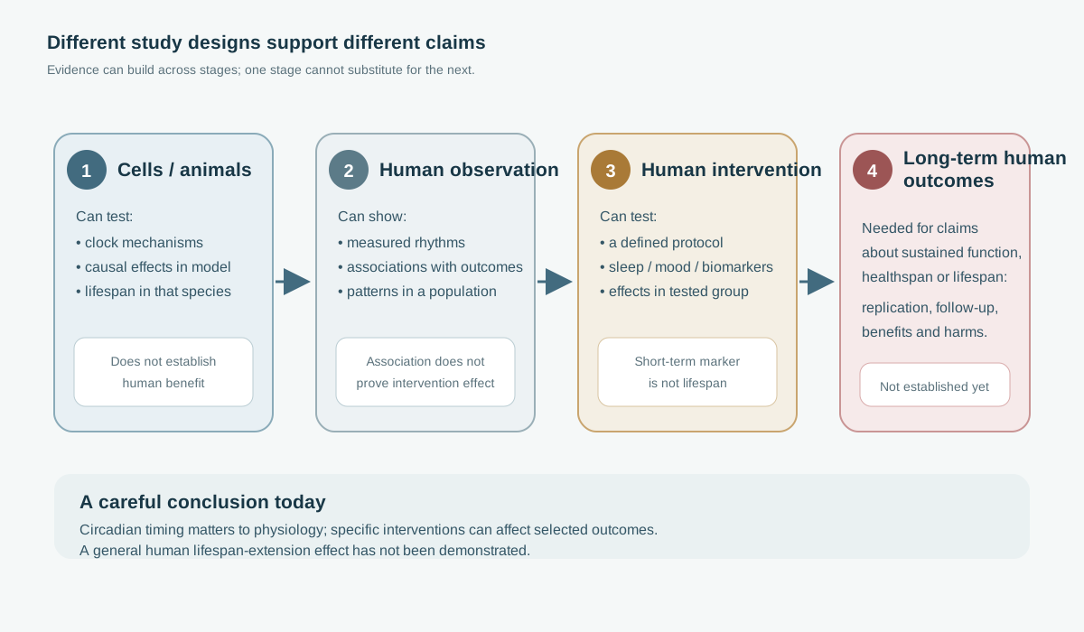
Until such evidence exists, a careful conclusion is narrower: circadian timing is part of human physiology; several rhythms and behaviors change with age in some contexts; and timed interventions can influence selected outcomes. It remains unknown whether deliberately strengthening or realigning rhythms slows biological aging or extends human lifespan.
- Measure rhythm features separately from sleep and activity.
- Test a defined intervention against an appropriate control.
- Follow meaningful health outcomes long enough to assess durability.
| Evidence level | What it can support | What it does not prove by itself |
|---|---|---|
| Cell / animal mechanism | Biological plausibility and causal effects in the tested model | Clinical benefit or longer life in humans |
| Human observational rhythm study | Association between a measured pattern and a health outcome | That changing the pattern will change the outcome |
| Human intervention trial | Effect of a defined protocol on measured outcomes in a stated group | Broad geroprotection if the outcome is short-term sleep, mood or a biomarker |
| Long-term replicated clinical evidence | Potential healthspan benefit when outcomes, population and harms are well characterized | Universal benefit beyond the populations and conditions studied |
For more context, see the biology hub, the longevity research overview, and the site’s biological-age measurement guide. Compare that with the biological clock review. These topics overlap, but circadian phase, sleep quality and biological-age algorithms should remain distinct in both research and personal interpretation.