
Normal brain aging versus neurodegenerative disease
Normal aging can involve slower processing, changes in sleep, altered network efficiency, modest volume loss and reduced physiological reserve. These changes vary widely and do not automatically indicate Alzheimer’s disease, mild cognitive impairment or another neurodegenerative disorder.
Neurodegenerative diseases have disease-specific pathology, clinical patterns and biomarker profiles. Aging can increase vulnerability, and disease can accelerate measured brain changes, but the two concepts should not be collapsed. A brain-age estimate is not a substitute for cognitive history, examination, fluid biomarkers, PET, or other validated assessment when disease is suspected.
Gray matter, white matter, connectivity and vascular function
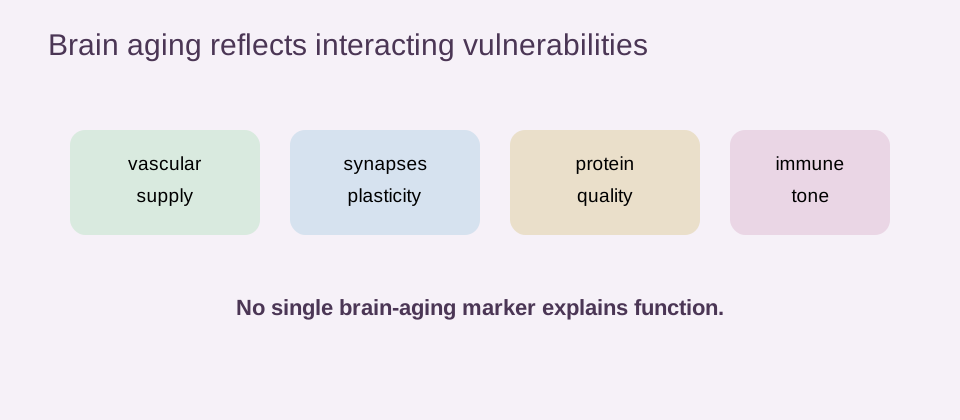
Across adulthood, gray-matter volume and cortical thickness can change, white-matter integrity can decline, and long-range network communication can become less efficient. The pattern is regional rather than uniform. Some systems remain resilient while others are more sensitive to vascular or metabolic stress.
Small-vessel disease, blood-pressure exposure, atherosclerotic risk, sleep disruption and impaired neurovascular coupling can affect both structure and function. These factors create shared pathways between cardiovascular health and cognition, but a population association does not identify the dominant pathway in one person.
| Mechanism | Possible signal | Evidence boundary |
|---|---|---|
| Vascular | white-matter change, perfusion or blood-pressure associations | not a stand-alone dementia diagnosis |
| Metabolic | glucose homeostasis and energy-use changes | transition patterns need replication |
| Immune | microglial, astrocytic and inflammatory signaling | activation is context-dependent |
| Synaptic | connectivity, plasticity and network efficiency | imaging proxy is not direct synapse count |
Synapses, plasticity and network efficiency
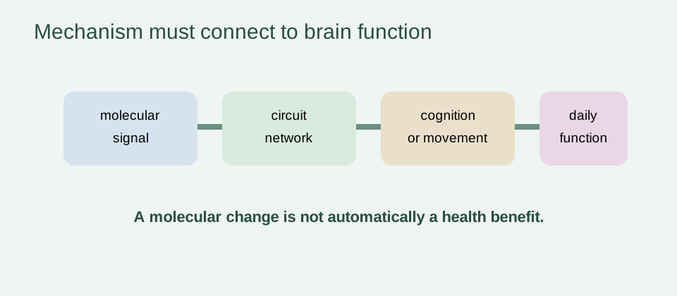
Synapses remain adaptable throughout life, but plasticity can be affected by sleep, stress, vascular supply, sensory input and metabolic state. Networks may compensate for local structural change by recruiting alternate pathways, so similar MRI findings can coexist with different cognitive performance.
Functional resilience is therefore a key companion to structure. Memory testing, executive function, processing speed and daily functioning measure related but non-identical outcomes. A model that predicts chronological age from anatomy should not be assumed to measure learning capacity or independence.
Mitochondrial, metabolic and glucose-related brain stress
Neurons require high and continuous energy supply. Mitochondrial dysfunction, altered glucose transport, oxidative stress and impaired waste handling can reduce reserve. The mitochondrial dysfunction page provides wider context, while this page keeps mechanistic plausibility separate from clinical brain rejuvenation.
A 2025 PNAS study analyzed multimodal data from about 19,300 participants and reported nonlinear brain-aging transitions with a midlife metabolic and glucose-homeostasis framing. Such a result can identify a window for research, but it does not establish a universal transition age or prove that a glucose intervention reverses brain aging.
Neuroinflammation, glia and the blood–brain barrier
Microglia and astrocytes support surveillance, repair and synaptic regulation. With age, their signaling environment can change, and systemic inflammation can influence the brain through vascular, endocrine and immune routes. The result may be impaired resolution, but immune activation is not uniformly harmful or uniformly reversible.
Blood–brain-barrier integrity and neurovascular function also affect transport, clearance and tissue response. The inflammaging page and immune-aging page describe why a lower inflammatory marker is not equivalent to restored neural function. Timing, cell type, tissue and clinical outcome all matter.
Protein homeostasis, autophagy and aggregation vulnerability
Protein quality control includes folding, trafficking, autophagy and clearance. Age-related stress can make neurons more vulnerable to misfolded or aggregated proteins, while disease-specific pathology can amplify those vulnerabilities. The proteostasis-aging page distinguishes a biological mechanism from a proven therapy.
Imaging, fluid biomarkers and post-mortem pathology measure different layers of this process. A structural brain-age model can correlate with disease risk without identifying amyloid, tau or another specific pathology. Disease-specific testing is required for disease-specific conclusions.
How MRI-derived brain-age models work
Researchers train a supervised model on MRI scans from a reference lifespan sample with known chronological ages. T1-weighted images, gray- and white-matter segmentations, cortical features, volumes or other derived measures may be used. The model is then applied to unseen scans to estimate brain-predicted age.
The brain-age gap is the difference between predicted and chronological age, often residualized for age, sex, scanner site, intracranial volume or other covariates. Bias correction and scanner harmonization matter because model error can vary across age groups, sites and acquisition protocols.
In one large 2025 genome-wide analysis, T1-weighted MRI and supervised models were used to estimate gray-matter, white-matter and combined brain age. The discovery sample reached a mean absolute error of about 3.09 years and a correlation of 0.86, while test–retest reliability for the residualized gap was high in the reported subsets.
These performance metrics describe estimation and repeatability. They do not establish that a score distinguishes normal aging from disease in a clinic, nor that it predicts benefit from an intervention. Scanner site, acquisition, preprocessing and reference population remain part of the model’s identity.
Brain age gap: what it measures and what it does not
A positive gap means the model output is higher than expected for the reference group. It may capture a mixture of normal variation, vascular risk, metabolic disease, smoking, alcohol, socioeconomic exposure, medication, image quality and prodromal disease. It does not isolate a causal aging rate.
Model reliability and predictive validity are separate. A score can be repeatable but weakly useful for an outcome, or predictive in one cohort but poorly calibrated in another. The organ-specific biological-age page explains why a brain score should remain attached to its modality, cohort and endpoint.
Regression toward the mean also matters. People selected because they have an extreme score may move closer to the reference average on repeat testing even without a biological change. A reported score should therefore include uncertainty, repeatability and the direction of the comparison.
For research use, the brain-age gap can be a compact phenotype for testing associations. For clinical use, it would need validated thresholds, demonstrated incremental value and evidence that acting on the result improves cognition, function or quality of life.
Genetics, APOE, cardiovascular risk and metabolic health
A 2025 Nature Aging genome-wide analysis used up to 56,348 individuals and reported 59 associated loci, with common variants explaining roughly 23–29% of brain-age-gap variance. It highlighted loci and pathways involving neurogenesis, immune processes and the MAPT region, while genetic correlations included mental health, diabetes, alcohol intake and socioeconomic traits.
The study also used Mendelian-randomization analyses that supported causal roles for high blood pressure and type 2 diabetes in accelerated brain aging under the assumptions of that method. This is stronger than a simple correlation but still does not create a treatment prescription or prove that lowering a brain-age score improves cognition.
Longitudinal evidence before cognitive impairment
Cross-sectional studies compare people at one time; longitudinal studies ask whether a baseline score predicts what happens later. A 2026 BrainAGE study reported that elevated MRI-derived brain age preceded later cognitive impairment and improved prediction of future cognitive decline in its cohort. A separate 2025 12-year study adds preclinical prediction context.
Prediction before diagnosis is clinically interesting, but it can still reflect early disease biology rather than an independent aging mechanism. The useful question is whether the score adds calibrated information beyond cognitive testing, vascular risk, education, genetics and established biomarkers, and whether that information changes an outcome.
Brain age versus cognition, fluid biomarkers and PET
Cognitive testing measures performance; MRI brain age summarizes structure or derived imaging features; fluid biomarkers can measure disease-related molecules; PET can visualize selected molecular or metabolic patterns. They can be complementary because they answer different questions.
A brain-age gap should not be used to infer amyloid or tau status, and a normal cognitive score does not exclude every pathology. Combining modalities can improve characterization, but added tests also increase complexity, cost and the risk of false-positive interpretation.
Which brain-aging factors are modifiable in humans?
Blood-pressure control, diabetes prevention or management, physical activity, sleep quality, smoking cessation, hearing and social engagement are relevant to brain health and disease risk. Evidence for reducing risk is not the same as evidence for reversing a validated brain-age biomarker or extending lifespan by doing so.
Exercise and metabolic interventions can improve vascular and functional outcomes in appropriate contexts. A study that changes cognition, MRI features or a brain-age score should report the design, adherence, comparator, duration and adverse events. One positive biomarker result cannot establish broad rejuvenation.
Drug and regenerative strategies under study
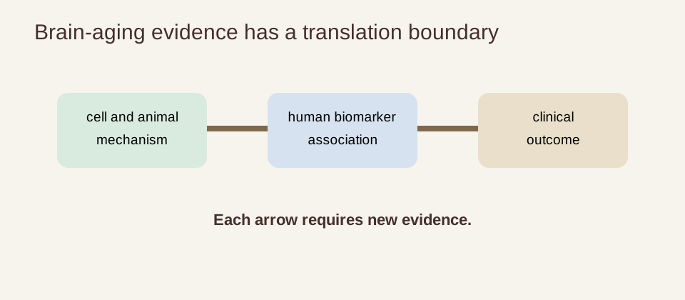
Research includes anti-inflammatory, metabolic, vascular, senolytic, proteostasis, regenerative and neuroplasticity-oriented strategies. Animal studies can identify mechanisms and improve performance under experimental conditions, but they cannot establish human cognitive benefit or lifespan extension.
Human drug trials need a defined population, clinically meaningful endpoints, adequate follow-up and safety monitoring. A treatment that changes a surrogate can also cause infection, vascular, psychiatric or other harms. No broad regenerative protocol has established reversal of human brain aging.
Even when a therapy improves a neural measure, the claim must remain tied to the measure that moved. Better perfusion, altered inflammatory signaling, a changed MRI feature, improved memory and reduced dementia incidence are distinct endpoints with different evidentiary requirements.
Long-term prevention trials are difficult because cognition changes slowly and outcomes are influenced by education, hearing, vascular disease, sleep, mood and social environment. Those difficulties increase the value of careful trial design; they do not justify substituting a convenient biomarker for a meaningful outcome.
Why a younger predicted brain age is not proof of reversed neurodegeneration
Models can be affected by scanner, preprocessing, age distribution and the features selected during training. An intervention may alter hydration, vascular tone, inflammation or image appearance without reversing disease pathology. Regression to the mean can also make an extreme baseline score move toward the average.
The strongest future studies will prespecify the brain-age model, validate it across sites, include cognitive and functional outcomes, track disease-specific biomarkers where relevant, and monitor harms. They will report null findings and duration rather than selecting only favorable biomarker changes.
Evidence verdict and clinical validation
Brain aging is a systems process involving structure, connectivity, vascular function, metabolism, immunity, proteostasis and reserve. MRI-derived brain-age gaps are increasingly reproducible and can predict later impairment or mortality-related outcomes in cohorts. Genetic and metabolic studies add plausible contributors and testable pathways.
The boundaries remain decisive. Normal aging is not dementia, an age gap is not a diagnosis, longitudinal prediction is not causation, and risk-factor modification is not proof of brain rejuvenation. No intervention has broadly reversed human brain age and extended human lifespan.
The responsible conclusion is brain-age models are promising research biomarkers whose clinical role remains under validation. A clinically useful test would need reliable measurement, transportable calibration, incremental value beyond existing assessment, a defined decision and evidence that using it improves outcomes without unacceptable harm.
- state whether evidence is cross-sectional, longitudinal, interventional or mechanistic;
- separate normal aging from neurodegenerative disease;
- keep MRI age gaps distinct from fluid biomarkers and PET;
- do not infer treatment need from a model output;
- require cognitive, functional and safety outcomes before claiming rejuvenation.