Why one biological age can be misleading
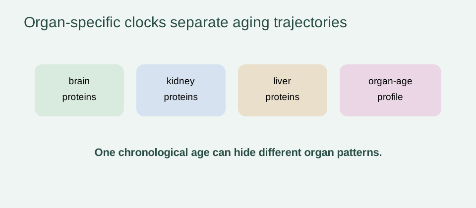
Chronological age is shared by everyone born in the same year, but tissues experience different exposures, workloads, injuries, infections and repair demands. Vascular risk can accumulate while cognition remains stable; kidney filtration can change for reasons that do not mirror brain aging; and immune remodeling can reflect exposure history as much as time.
A whole-body biological-age score compresses that variation into one number. Compression can be useful for a broad risk model, but it can also hide the organ or pathway most relevant to a person’s outcome. Organ-specific models attempt to preserve some of that heterogeneity.
This does not mean that an organ score is automatically more truthful. It means the question has been narrowed: which measured features are associated with the aging pattern of a named system, and do they add useful information beyond overall age and standard clinical risk?
Chronological age versus organ-specific biological aging
An organ-age estimate is usually predicted from biomarkers or images and compared with chronological age. The difference is an age gap: positive values indicate that the model’s output is higher than expected for the reference population, while negative values indicate the opposite. The direction is model-relative, not a universal unit.
The biological-age testing page explains why different clocks can disagree. Proteomic, MRI, clinical and methylation clocks sample different layers and time scales. Agreement may support a shared signal, but disagreement is not proof that one organ is objectively older.
| Result | Reasonable interpretation | Overreach to avoid |
|---|---|---|
| Positive age gap | model output is higher than expected for the reference group | the organ is literally that many years old |
| Disease association | score is statistically related to an outcome | the score caused the disease |
| Organ specificity | signal is enriched for a nominated system | confirmed pathology in that organ |
| Change after treatment | measurement moved in a study | the organ was rejuvenated or lifespan extended |
How organ-age models are built
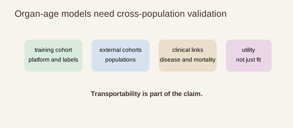
Researchers select an input modality, define a target such as chronological age, and train a model in a development cohort. They may use feature selection, regularization or nonlinear machine learning. A credible report keeps test data separate from training and tuning, then evaluates calibration and performance in an external cohort.
Organ enrichment can be inferred from tissue expression references, while MRI models can use anatomical or functional imaging features. The output is not a direct biopsy or a complete physiological examination. It is a model-based summary of the features the investigators chose to measure.
Training choices also shape the age gap. Age range, sex distribution, missing-data rules, image quality, protein normalization and covariate adjustment affect what the model learns. A clock that performs well in a narrow research cohort may need recalibration before it is used in a different population.
External validation should report more than a single correlation. Calibration, error across age groups, performance in people with disease, and incremental value beyond routine clinical measures determine whether the score is useful for a particular purpose.
Proteomic organ clocks and MRI-based organ clocks
Proteomic clocks use circulating proteins that may reflect secretion, inflammation, metabolism, injury or clearance. MRI clocks use image-derived features and can provide structural or functional information at the organ level. The two modalities can complement one another, but their age gaps are not interchangeable.
The 2025 Nature Medicine MRI study described multi-organ clocks in 313,645 individuals and integrated imaging-derived age gaps with proteins, metabolites and genetics. Its scale shows the potential of imaging-based systems phenotyping; it does not turn an MRI age gap into a routine diagnosis.
Independent plasma-model work has also framed disease as accelerated aging of organismal systems. Such models can be useful for comparing organ contributions and for asking whether one score adds predictive information beyond another. Their statistical framing still leaves open whether the signal is a cause, a consequence or a mixture of both.
Modality changes the practical meaning of the result. A protein-based score may respond to circulating physiology before structural change is visible, while an MRI score may capture anatomy that is less sensitive to a short-lived inflammatory episode. Neither is automatically closer to the truth; validity depends on the outcome being predicted and the decision being supported.
Brain, cardiovascular and arterial aging signals
Brain age scores can capture a mixture of vascular, structural, metabolic and neurobiological features. In the large 2025 proteomic study, brain aging showed the strongest association with all-cause mortality among the organ scores and was linked with neurodegenerative outcomes. That is a population-level association, not a diagnosis of dementia in an individual.
Cardiovascular and arterial scores can reflect vascular remodeling, lipid-related biology, blood pressure, inflammation and cardiac stress. A higher score may be associated with cardiovascular disease while still being influenced by shared exposures that affect several organs. Clinical history, examination and validated tests remain the decision-making layer.
Kidney, liver, metabolic and immune-system aging
Kidney-related scores are particularly sensitive to filtration, metabolic state and medication context. Liver-related signals can reflect protein synthesis, clearance, metabolic stress or injury. Metabolic organs are connected through circulation, so an organ label should be treated as an evidence-weighted hypothesis rather than an isolated cause.
Immune aging is a system-level phenotype involving hematopoiesis, lymphocytes, innate cells and inflammatory signaling. The immune-aging page explains why an immune score cannot be reduced to one cell count or used to justify indiscriminate stimulation.
Cross-organ coupling and the 2025 proteomic study
Organs do not age in isolation. Shared blood flow, endocrine signaling, inflammation, behavior and disease create coupling, while partially independent trajectories can still be detected. The practical goal is not to rank organs from “oldest” to “youngest,” but to understand which patterns add information for a defined outcome.
The 2025 Nature Aging study developed an organismal clock and ten organ-specific clocks in 43,616 UK Biobank participants, with validation in 3,977 China Kadoorie Biobank participants and 800 Nurses’ Health Study participants. It used an Olink Explore 3072 panel measuring 2,916 proteins, with tissue-enrichment information from GTEx.
In that study, organ-specific age gaps were generally only weakly correlated with one another, suggesting some partial independence. All organ gaps were associated with mortality, with the brain showing the strongest association in the reported analyses. These findings support a multiorgan view of risk, not a ranking that can be applied directly to an individual.
The study also illustrates why the cohort and adjustment set belong beside every headline result. The analyses adjusted for demographic and lifestyle variables and were replicated across cohorts, but the models were still developed from observational data. Replication strengthens confidence in the association; it does not remove all confounding or establish that changing the score changes survival.
Multi-tissue atlases, age trajectories and inflection points
A 2025 Cell study analyzed 516 samples from 13 human tissues spanning five decades. It reported tissue-specific proteomic age clocks and an aging inflection around age 50, with blood vessels described as an early and particularly susceptible tissue in that dataset. These findings map trajectories and candidate mechanisms; they do not specify when an individual’s organ will fail.
Cross-sectional age curves can be shaped by sampling, survivor bias, cohort composition and disease. Longitudinal data are better suited to individual change but still require careful handling of assay drift, regression to the mean and changing medication or health status.
An atlas can also reveal transcriptome-proteome decoupling, proteostasis decline and tissue-specific protein changes that are not visible in a single blood measurement. Those observations help connect organ-age scores to mechanisms such as vascular remodeling or altered protein quality control. They remain observational evidence until perturbation and clinical outcome studies test the proposed pathway.
Inflection points should therefore be reported as features of a dataset and model, not as universal biological deadlines. A curve that changes near age 50 can guide sampling or hypothesis design, but it cannot establish that everyone should begin a supplement, scan or intervention at that age.
Genetics, environment and reverse causation
Organ-age gaps can reflect inherited variation, lifestyle, sleep, smoking, physical activity, diet, socioeconomic exposure, infection and chronic disease. Genetic associations can suggest pathways, but they do not prove that changing a protein or gene will rejuvenate an organ.
Reverse causation is a persistent concern: an established disease or its treatment can make an organ score appear older. A prospective association before diagnosis is more informative than a cross-sectional difference, but even prospective prediction can include prodromal disease rather than an independent aging mechanism.
Can interventions selectively change one organ-age clock?
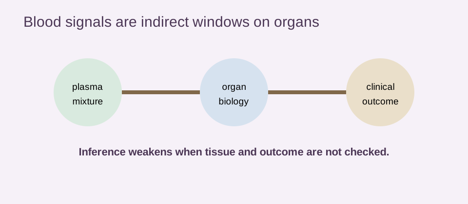
In principle, a treatment could affect one organ more than another. In practice, a changed score may reflect systemic inflammation, weight, blood pressure, medication, fluid balance or assay variation rather than selective rejuvenation. The intervention, platform, sampling interval and prespecified clock must all be reported.
A strong trial would randomize where feasible, include a comparator, repeat the measurement, monitor harms and measure a clinical or functional endpoint. The longevity clinical trials page covers why a biomarker response is not automatically a surrogate for healthspan or survival.
Why consumers should not interpret organ age as a diagnosis
Consumer reports often present an age gap with a reassuringly precise number. Precision in the display does not guarantee precision in the underlying biology. Without the model, assay, reference population, calibration and clinical validation, the number cannot establish disease or tell someone what action to take.
The proteomic-aging-clocks page describes platform and cohort limits, while the brain-aging page provides organ-specific context. An unusual result should be discussed in the context of symptoms and standard medical evaluation, not treated as a diagnosis or a reason to replace indicated care.
Evidence verdict and path toward clinical validation
Human evidence supports the idea that aging is heterogeneous across organs. Proteomic and MRI studies can estimate partially independent organ-related patterns, and accelerated scores can associate with future disease and mortality. Multi-tissue atlases provide biological context and identify tissue-specific trajectories.
The evidence does not support treating an age gap as a literal organ age, diagnosis or proven intervention target. Modality, cohort, calibration, platform drift, confounding, reverse causation and clinical endpoint validity all matter. Improving one score would not prove whole-body rejuvenation.
The responsible conclusion is organ-specific biological-age clocks are promising research tools whose clinical meaning remains under validation. The next step is prospective testing showing that a specified score improves a real decision or outcome, with durable benefit and acceptable risk.
For readers, the most durable takeaway is methodological: keep the organ label, modality, cohort and endpoint attached to the result. That discipline makes the page useful when future studies refine, recalibrate or contradict today’s clock estimates. It also reduces false precision when comparing commercial and academic reports across populations.
- name the modality, cohort and reference population;
- separate cross-sectional association from prospective prediction;
- treat organ enrichment as context, not a diagnosis;
- compare scores with validated clinical measures;
- require intervention and outcome evidence before claiming rejuvenation.