Biology of aging · evidence reviewed September 2026
What mitochondria do beyond ATP production
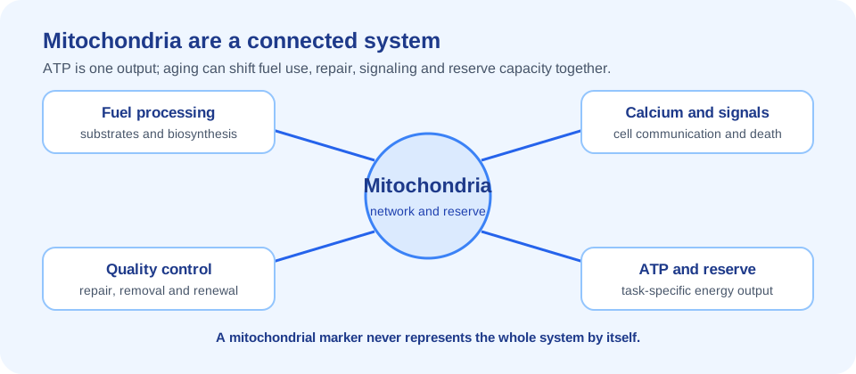
Mitochondria are membrane-bound organelles that coordinate far more than ATP production. They process fuels, contribute to biosynthesis, regulate calcium, participate in cell death, communicate stress to the nucleus and shape innate immune signaling. In the brain, heart and skeletal muscle, their ability to match energy supply with changing demand is central to tissue function.
ATP is an important output, not the whole system. A cell can maintain ATP temporarily by changing fuel use, reducing demand or recruiting reserve capacity while other defects accumulate. Conversely, a respiratory measurement can change without causing an immediate symptom. The meaning of a mitochondrial result depends on tissue, task, substrate, temperature, activity and disease context.
A modern mitochondrial-aging model asks whether the organelle population is being maintained. Damaged proteins and membranes must be repaired or removed; mitochondrial networks must divide and fuse; new organelles must be produced; and signals from stressed mitochondria must be contained. The parent Biology of Aging hub places these processes beside other hallmarks rather than treating one pathway as the master explanation.
Why mitochondrial dysfunction is a hallmark of aging
Mitochondrial dysfunction is included in modern hallmarks-of-aging frameworks because mitochondrial changes interact with cellular senescence, nutrient sensing, proteostasis, stem-cell function and chronic inflammation. The relationship is bidirectional. Aging processes can impair mitochondria, while mitochondrial stress can change cell behavior and tissue communication.
The phrase “dysfunction” covers several possible observations: lower respiratory capacity, altered substrate oxidation, membrane-potential changes, abnormal morphology, mtDNA mutations, impaired mitophagy, defective proteostasis or excessive inflammatory signaling. These are not interchangeable. A study that measures one should not claim to have measured all mitochondrial aging.
Human relevance is strongest when mitochondrial measures connect to function. Skeletal-muscle mitochondrial energetics, for example, may help explain mobility decline in older adults, but the association does not prove that correcting one measurement prevents disability. Mitochondrial biology is a plausible bridge between molecular aging and function, not a shortcut around clinical evidence.
Mitochondrial DNA damage, heteroplasmy and clonal expansion
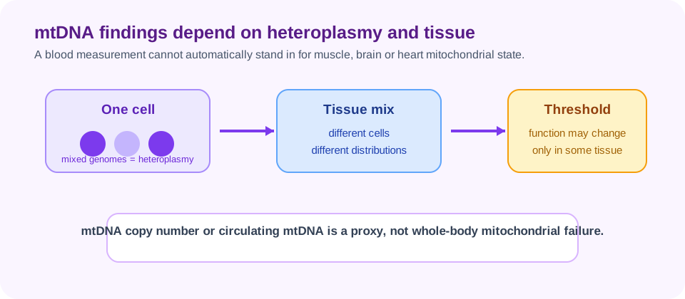
Mitochondrial DNA, or mtDNA, sits close to the respiratory machinery and is copied separately from nuclear DNA. Variants can arise through replication errors, damage or inherited transmission. Cells may contain a mixture of mitochondrial genomes, a state called heteroplasmy. The fraction and distribution of a variant can influence whether a tissue crosses a functional threshold.
Age-related mtDNA changes are not uniform across people or organs. A blood measurement may not represent skeletal muscle, brain or heart. Clonal expansion can make some variants more prominent in a cell or tissue, while selection, turnover and mitochondrial quality control influence which genomes persist.
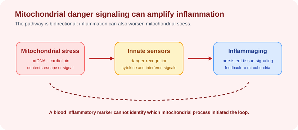
mtDNA damage is also a signaling issue. When mitochondrial contents escape into the cytosol or circulation, they can be interpreted as danger signals and stimulate innate immunity. This is one route by which mitochondrial stress can connect to inflammaging, but the presence of an inflammatory signal does not prove that mtDNA damage is its only cause.
| Measurement | What it indicates | What it does not establish alone |
|---|---|---|
| Heteroplasmy | Mixture of mitochondrial genome variants. | Whole-body mitochondrial failure. |
| mtDNA copy number | A proxy influenced by cell type and mitochondrial content. | Respiratory capacity or tissue function by itself. |
| Circulating mtDNA | Possible stress or cell-injury signal. | A single cause of systemic inflammation. |
| Muscle mtDNA assay | Genetic state in a sampled tissue. | Brain, heart or immune-cell mitochondrial status. |
Electron transport and bioenergetic changes
The electron-transport chain transfers electrons through respiratory complexes and uses the resulting energy gradient to support ATP synthesis. Aging, inactivity, disease and environmental stress can alter complex activity, substrate use, membrane potential and reserve capacity. A person may have enough energy for quiet conditions but less capacity during exercise, infection or repair.
Bioenergetic measurements depend heavily on protocol. Oxygen consumption can be assessed in isolated mitochondria, permeabilized fibers, cells or intact organisms, each answering a different question. The substrate supplied, temperature, oxygen level, normalization method and physical activity before sampling can change the result.
A lower measurement can be a cause, consequence or adaptation. For example, reduced activity may lower the demand that maintains oxidative capacity, while disease can damage both muscle and mitochondria. This is why the relationship between mitochondrial energetics and mobility deserves longitudinal study rather than a one-time “energy age” label.
- Capacity: maximum respiration under a defined protocol.
- Reserve: ability to increase output above baseline demand.
- Efficiency: relationship between fuel oxidation, gradient and ATP production.
- Adaptation: response to exercise, fasting, illness or another stress.
Reactive oxygen species: signal versus damage
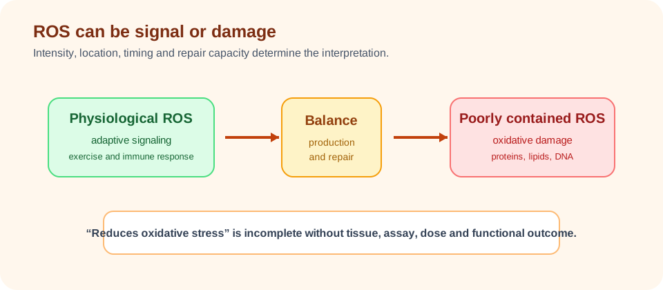
Reactive oxygen species, or ROS, are produced during normal metabolism and can act as signals that adjust antioxidant defenses, inflammation and adaptation. Excessive or poorly contained ROS can oxidize proteins, lipids and nucleic acids. The key question is not whether ROS exist, but whether production, detoxification, repair and signaling remain balanced in a particular tissue.
This distinction makes blanket antioxidant claims unreliable. Suppressing every ROS signal could interfere with exercise adaptation or immune function, while ignoring sustained oxidative damage could miss a contributor to disease. The effect of a compound depends on dose, timing, tissue exposure, baseline redox state and the endpoint measured.
Mitochondrial glutathione, superoxide handling, lipid oxidation and redox-sensitive transcription are related but distinct measures. A study should report which one changed and whether the change connected to function. “Reduces oxidative stress” is too vague to carry a broad longevity conclusion.
Mitochondrial quality control: proteostasis, dynamics, mitophagy and biogenesis
The 2026 human-longevity framework treats mitochondrial quality control as an integrated system. It includes proteostasis, mitochondrial dynamics, removal of damaged organelles, production of new organelles and repair-by-replacement processes. Failure in one module can increase pressure on the others, but the modules are not redundant and cannot be summarized by one “mitochondrial score.”
Proteostasis and the mitochondrial stress response
Proteostasis keeps the organelle’s protein machinery usable. Mitochondria import many proteins made in the cytosol and must fold, assemble and remove them under changing conditions. Proteases, chaperones and stress-response pathways help maintain function. The mitochondrial unfolded-protein response and integrated stress response can be adaptive when brief, but persistent stress may alter metabolism and inflammatory behavior.
Fission, fusion and cristae remodeling
Fusion can mix contents and support network resilience, while fission can isolate damaged segments and prepare them for removal. Cristae structure shapes respiratory-chain organization and can influence how efficiently electrons and protons are handled. More fusion or more fission is not universally better; the appropriate balance depends on tissue and stress.
Mitophagy and removal
Mitophagy is the selective delivery of damaged mitochondria to lysosomal degradation pathways. PINK1, Parkin and receptor-mediated routes are among the systems involved, but mitophagy is not a single switch. Flux, cargo selection and lysosomal capacity matter. The dedicated mitophagy and aging page follows this mechanism in greater detail.
Biogenesis and renewal
Biogenesis replaces mitochondrial mass through coordinated nuclear and mitochondrial gene expression. Exercise, nutrient sensing, AMPK, PGC-1-family signaling and other inputs can influence the process. Producing more mitochondria is not automatically beneficial if quality control, assembly or fuel delivery remains impaired.
Mitochondrial-derived vesicles and repair
Mitochondrial-derived vesicles can selectively transport damaged or oxidized cargo to other cellular compartments. This provides a repair and disposal route that complements whole-organelle mitophagy. It also shows why the word “quality control” includes selective trafficking, not only degradation.
These pathways are active areas of research, especially in the interface between mitochondria and the endoplasmic reticulum. Their presence in a model system does not establish that a supplement or exercise protocol can safely manipulate them in humans.
mtDNA release, innate immunity and inflammaging
When mitochondria are damaged or stressed, mtDNA, cardiolipin and other mitochondrial products can activate innate immune sensors. The resulting cytokine and interferon responses may help contain injury, but persistent signaling can contribute to chronic inflammation. This is one mechanistic route linking mitochondrial dysfunction to altered intercellular communication.
Inflammation can also damage mitochondria, creating a feedback loop. Infection, obesity, tissue injury and senescence can all change the local environment. A blood inflammatory marker cannot identify which mitochondrial process initiated the signal, so causal claims require tissue and longitudinal evidence.
The clinical implication is not that every inflammatory condition needs a “mitochondrial supplement.” It is that interventions should be evaluated against a defined disease or function outcome, with biomarkers used to explain or monitor the response.
Mitochondria in stem-cell aging and immune aging
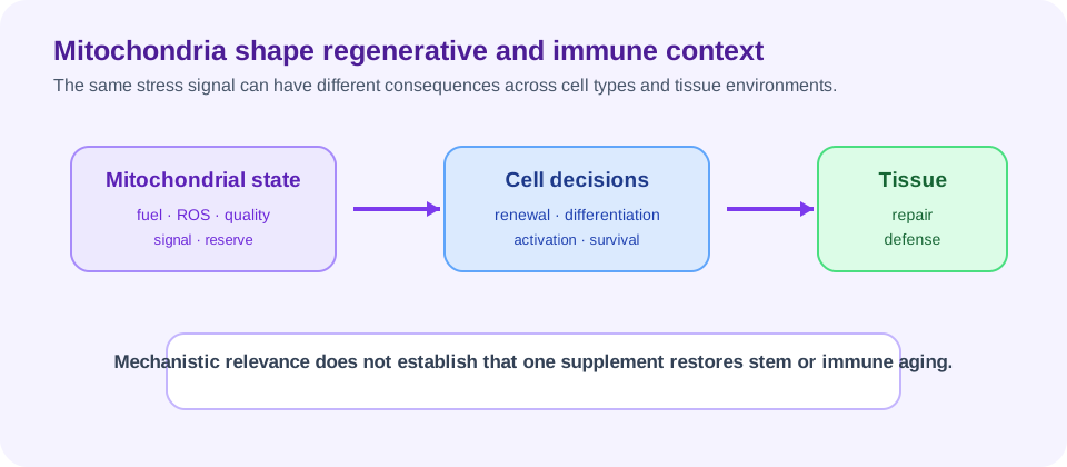
Stem cells must balance energy production, biosynthesis, self-renewal and differentiation. Mitochondrial metabolism can influence whether a stem cell remains quiescent or enters a differentiation program. With age, altered quality control, redox signaling and inflammatory exposure may reduce regenerative resilience, but the direction and mechanism differ by stem-cell compartment.
Immune cells also reprogram mitochondrial metabolism as they activate, migrate and form memory. Age-related immune dysfunction may involve impaired mitochondrial quality control, altered autophagy and chronic inflammatory signaling. A mitochondrial measurement in muscle should not be assumed to represent immune-cell aging.
- Stem-cell status depends on tissue niche and differentiation state.
- Immune-cell metabolism changes with activation and infection history.
- Circulating biomarkers average signals from many tissues.
- Regeneration and immunity require context-specific endpoints.
Why mitochondrial defects differ by tissue
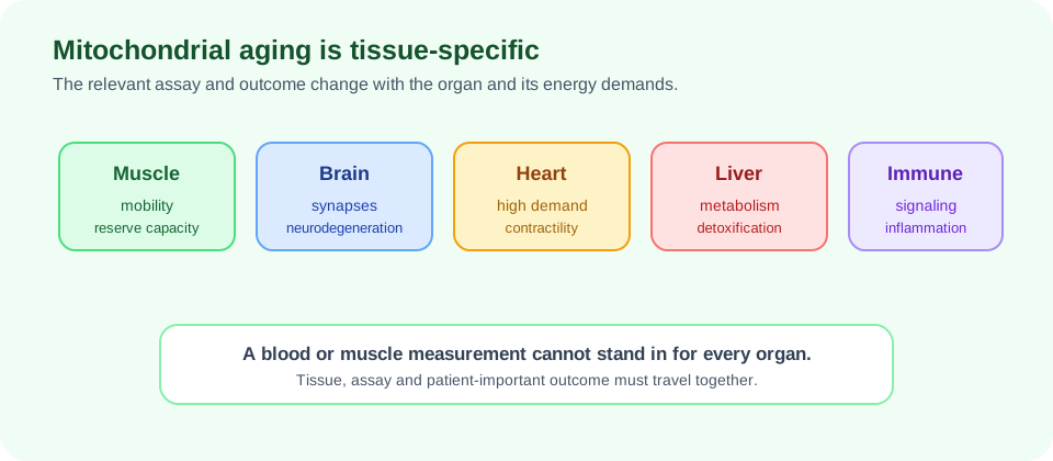
Mitochondria are not identical across organs. Heart and skeletal muscle have high energetic demand and specialized contractile workloads. Neurons must sustain long-lived axons and synapses. Liver mitochondria integrate nutrient and detoxification signals. Immune cells change metabolic programs rapidly. Kidney, retina, skin and reproductive tissues have their own exposure, turnover and quality-control constraints.
Age-related decline can therefore be patchy. A person may have reduced muscle reserve while maintaining other mitochondrial functions, or a blood assay may look stable while a vulnerable neural circuit changes. This tissue heterogeneity is one reason whole-body biological-age claims from a single mitochondrial biomarker require caution.
It also changes intervention design. A treatment that reaches muscle may not reach the brain. Exercise can affect many tissues but with different timing and dose-response. A mitochondrial intervention should specify its target organ, delivery, mechanism and clinically meaningful outcome.
How mitochondrial function is measured in humans
Human mitochondrial studies use a wide range of measurements: oxygen consumption in muscle fibers, magnetic-resonance spectroscopy, exercise response, metabolomics, mtDNA features, circulating proteins, redox markers and tissue imaging. Each offers a partial view. Some are direct functional assays; others are proxies that depend on multiple biological systems.
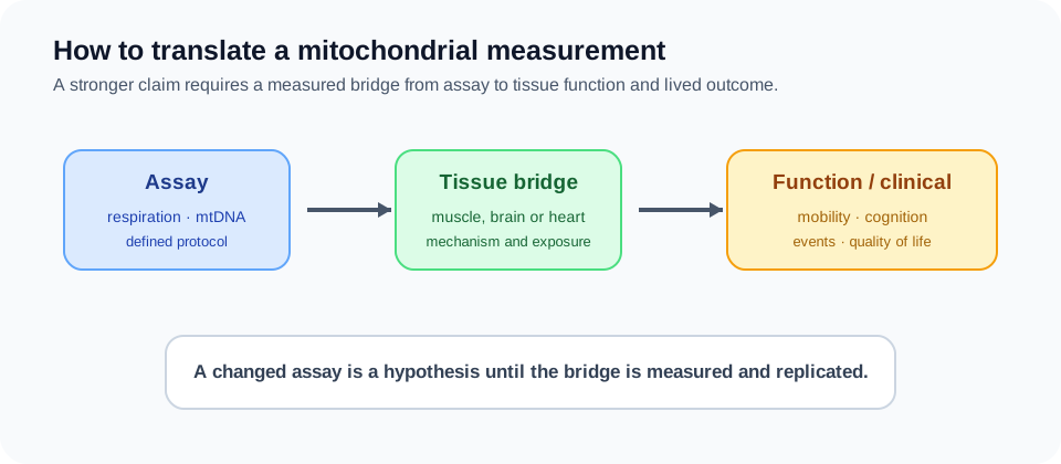
Measurement quality includes sampling site, fasting and exercise state, assay normalization and technical reproducibility. It also includes whether the result predicts a meaningful outcome. A single time point cannot show a trajectory. A surrogate can be informative without being validated as a treatment target.
The most persuasive human evidence combines a mechanistic measure with longitudinal function, disease outcomes or quality of life. For example, a muscle respiratory change becomes more relevant if it tracks reproducible mobility or exercise-capacity improvement. Even then, association and causation must be separated.
| Evidence layer | Example | Interpretive limit |
|---|---|---|
| Molecular | mtDNA, transcripts or redox marker. | May not reflect organelle performance. |
| Functional | Respiration or reserve capacity in sampled tissue. | May not represent another organ. |
| Physiological | Exercise response, glucose handling or cardiac function. | Many systems contribute to the result. |
| Clinical | Mobility, symptoms, events or quality of life. | Needs adequate duration and power. |
Interventions: what has reached humans
Exercise, adequate nutrition, sleep, disease treatment and activity remain the most established ways to influence whole-body physiology that supports mitochondrial function. They are not single-target “mitochondrial rejuvenation” therapies, and their effects depend on baseline health, dose, adherence and tissue.
Research has also examined NAD-related strategies, AMPK activators, caloric restriction, urolithin A, creatine, GlyNAC and other compounds. The evidence is intervention-specific. The NAD+ metabolism page, AMPK page, urolithin A page, creatine page and GlyNAC page keep their individual human evidence separate.
Preclinical improvement in mitophagy, respiration, membrane potential or lifespan is not a human clinical result. A supplement can change a biomarker without improving function, and a functional benefit can occur through several pathways without proving mitochondrial repair. Good trials state which claim they are actually testing.
- Define the target tissue and mitochondrial process.
- Use an appropriate comparator and prespecified primary endpoint.
- Measure safety and clinically meaningful function.
- Follow participants long enough to test durability.
Evidence verdict and key unsolved questions
Mitochondrial dysfunction in aging is a network problem involving quality control, dynamics, proteostasis, mitophagy, biogenesis, mtDNA integrity, redox signaling, immune communication and tissue-specific demand. The 2026 human-longevity literature emphasizes that repair, removal, remodeling and renewal operate together. This is a more useful model than treating ATP decline or oxidative stress as the entire story.
The strongest human conclusion is that mitochondrial measures are relevant to physiology and may help explain age-related functional decline. The weaker conclusion is that changing one mitochondrial marker will broadly reverse aging. No single mitochondria-targeted supplement or protocol has yet proved that claim.
Key unanswered questions include which mitochondrial defects are causal in each tissue, how baseline status changes response, whether blood markers can predict tissue function, how quality-control flux should be measured in humans and which interventions improve durable mobility, cognition, immune resilience or disease outcomes. Those questions require careful trials, not a single universal mitochondrial score.