What mitophagy is
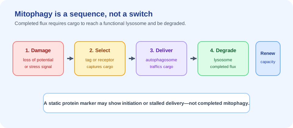
Mitophagy is the selective removal of mitochondria through autophagic machinery. A damaged or surplus mitochondrion is recognized, marked or captured, surrounded by an autophagic membrane, delivered to a lysosome and broken down. The components are coordinated with mitochondrial dynamics, trafficking, stress responses, replacement and cellular metabolism.
The word “selective” is essential. Basal mitophagy can help maintain a population of mitochondria without eliminating every organelle that experiences a transient fluctuation. A brief change in membrane potential during exercise or nutrient stress may be adaptive rather than a reason for wholesale removal. The cell must distinguish damage that can be repaired from cargo that should be retired.
Current mammalian biology is broader than the familiar PINK1/Parkin story. The 2026 review in Nature Reviews Molecular Cell Biology describes PINK1/Parkin-dependent and independent pathways, different triggers and tissue contexts. That breadth is why mitophagy belongs inside the wider mitochondrial dysfunction and aging framework rather than being treated as a standalone longevity lever.
Why damaged mitochondria must be selectively removed
Mitochondria generate energy, regulate redox chemistry, participate in calcium handling and communicate with innate immunity. A damaged organelle can therefore create more than an ATP shortfall. It may produce abnormal reactive oxygen species, release mitochondrial DNA or other danger signals, disrupt calcium balance, consume resources or interfere with neighboring mitochondria.
Selective removal can reduce the burden of dysfunctional cargo and make room for replacement. It can also shape how a cell adapts to exercise, starvation, infection, hypoxia or differentiation. But removal has costs: autophagy consumes membrane and lysosomal capacity, and excessive mitochondrial loss can leave a cell unable to meet energy or biosynthetic demands.
| Observation | Reasonable interpretation | Unsupported leap |
|---|---|---|
| More mitophagy-related protein | A pathway component or marker changed. | Completed mitochondrial clearance increased. |
| More mitochondrial fragments | Dynamics or stress changed. | Damaged mitochondria were successfully removed. |
| Higher lysosomal signal | Degradation capacity or cargo delivery may be involved. | Healthspan improved. |
| Improved muscle endpoint | A defined functional outcome changed. | Whole-body aging was slowed. |
The pathway map: PINK1/Parkin and other routes
PINK1/Parkin-dependent mitophagy
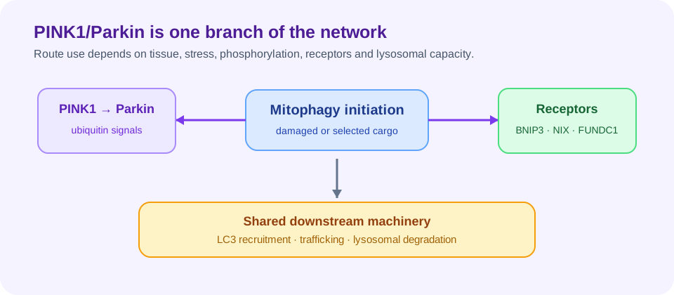
When mitochondrial import and membrane potential are sufficiently disrupted, PINK1 can accumulate on the outer mitochondrial membrane instead of being rapidly processed. PINK1 phosphorylates ubiquitin and helps activate the E3 ligase Parkin. Parkin then amplifies ubiquitin signals on outer-membrane proteins, recruiting autophagy receptors such as optineurin and NDP52 and associated TBK1 signaling.
This pathway is experimentally powerful and clinically relevant: recessive mutations in PINK1 and PRKN, the gene encoding Parkin, cause early-onset Parkinson’s disease. That genetic evidence demonstrates that the pathway matters in human disease, but it does not mean every age-related change in any tissue is caused by insufficient PINK1/Parkin activity.
Receptor-mediated and PINK1/Parkin-independent mitophagy
Other routes use mitochondrial outer-membrane receptors and their LC3-interacting regions to recruit autophagic machinery. BNIP3, NIX/BNIP3L and FUNDC1 are examples discussed in the mammalian literature, with activation shaped by hypoxia, phosphorylation, transcriptional state and cell type. Lipid signals and soluble receptors can also contribute.
The distinction is not merely a molecular catalogue. A pathway that dominates during red-blood-cell maturation, hypoxia or muscle remodeling may not be the same pathway operating in a neuron or an immune cell. The current mechanistic review emphasizes that the physiological roles and conditions in which these pathways operate are still being defined.
Dynamics, lysosomes and completed autophagic flux
Fission and fusion set the cargo context
Fission can separate a damaged segment from a healthier mitochondrial network, while fusion can mix contents and support resilience. These processes affect the size, connectivity and accessibility of potential cargo. Mitophagy proteins can influence or respond to mitochondrial dynamics, so a change in fragmentation is not itself a direct measure of clearance.
Cristae remodeling and contact with the endoplasmic reticulum also influence how mitochondria sense stress and interact with autophagic membranes. An intervention that changes morphology may be affecting network adaptation, mitophagy initiation, or both. Interpretation requires time-resolved measurements rather than a single microscopy image.
Lysosomal delivery is the point many assays miss
A mitophagy signal is incomplete if marked mitochondria accumulate because delivery or degradation is blocked. Autophagic flux asks whether material moves through the pathway over time. Researchers may combine reporters, lysosomal inhibition, microscopy, isotope tracing, proteomics or biochemical turnover assays to distinguish increased initiation from completed degradation.
This is the same conceptual problem described on the related autophagy and longevity page. Static LC3, Parkin, PINK1, BNIP3 or lysosome abundance can be informative, but each is a proxy with context-dependent meaning. “Higher marker expression” should not automatically be translated into “more healthy mitochondria removed.”
How mitophagy changes with aging
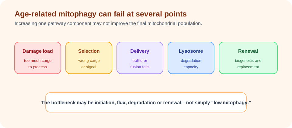
Aging can change mitochondrial damage load, lysosomal capacity, autophagosome formation, trafficking, nutrient sensing and the ability to replace lost organelles. The direction is not uniform across tissues or across experimental conditions. A pathway may be impaired in one compartment while adaptive signaling is preserved or increased in another.
The 2026 Nature Metabolism review on mitochondrial quality control in human aging places mitophagy alongside proteostasis, dynamics, biogenesis and repair/remodeling. This systems view matters because a bottleneck downstream of cargo selection can make stronger upstream signaling ineffective. If lysosomes cannot process the load, more initiation could increase congestion rather than restore quality.
Age-related disease studies often report associations between altered mitophagy markers and neurodegeneration, metabolic disease, inflammation or muscle decline. Association helps identify mechanisms, but it does not reveal whether the change initiated disease, responded to injury or represented a compensatory adaptation. Disease-specific evidence should not be relabeled as proof of a general anti-aging effect.
For readers following Biology of Aging, the practical conclusion is that mitophagy is a plausible control point, not a validated whole-body aging clock. The relevant outcome may be muscle reserve, neuronal resilience, immune function or another tissue-specific endpoint.
Inflammation, mtDNA release and tissue context
Damaged mitochondria can release mitochondrial DNA, cardiolipin and other molecules that activate innate immune sensors. Conversely, inflammatory cytokines, infection, senescence and metabolic stress can alter mitochondrial quality control. Mitophagy can therefore participate in feedback loops between organelle damage and inflammaging, but the presence of inflammation does not identify a single upstream defect.
Neurons face long axonal distances and limited replacement options. Skeletal muscle must adapt to repeated contraction and preserve reserve capacity. Liver cells integrate nutrient and detoxification signals, while immune cells remodel metabolism rapidly during activation and memory formation. The same PINK1, Parkin or lysosomal measurement can have different implications in each setting.
The 2026 npj Aging review on targeting mitophagy for neuroprotection illustrates why brain applications need their own evidence chain. A compound can be mechanistically attractive in a neuronal model yet fail to reach the relevant brain compartment, alter the wrong pathway or lack a validated human functional endpoint.
| Tissue | Distinct challenge | Outcome that would matter |
|---|---|---|
| Skeletal muscle | high workload, remodeling and reserve capacity. | strength, endurance, mobility or recovery. |
| Brain | long-lived neurons and axonal transport. | validated cognition, function or disease outcome. |
| Immune cells | rapid metabolic reprogramming during activation. | defined immune response or inflammatory disease endpoint. |
| Liver | nutrient sensing, detoxification and metabolic flux. | validated metabolic or disease outcome. |
How mitophagy is measured in living humans
Directly measuring completed mitophagy in a living person is difficult. A muscle biopsy can provide tissue-specific protein, transcript, imaging or ultrastructural information, but it samples a small region. Blood may contain circulating mitochondrial DNA, proteins or metabolites, yet those signals integrate multiple organs and may reflect injury, immune activity or clearance rather than mitophagy itself.
Researchers can study mitochondrial respiration, mitochondrial content, autophagy-related proteins, ubiquitination, lysosomal markers, isotope turnover, extracellular vesicles and imaging reporters. Each assay answers a narrower question than the phrase “mitophagy increased.” The strongest designs combine pathway measures with flux information and a prespecified physiological or clinical outcome.
Human evidence should also report timing. A brief stress response may be adaptive; a chronic marker elevation may reflect stalled degradation. Sampling after exercise, fasting, infection or medication can produce different values. Without repeated measures and clear normalization, a cross-sectional marker is a weak basis for an intervention claim.
Exercise, nutrition and pharmacological interventions
Exercise can alter mitochondrial turnover, energy demand, redox signaling and muscle remodeling. Nutrient stress and energy-sensing pathways can influence autophagy-related machinery. Pharmacological and nutritional candidates may target upstream signaling, receptor activity, mitochondrial stress, lysosomal function or replacement. These categories are biologically related but not interchangeable.
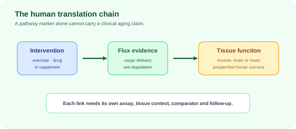
A convincing intervention trial would specify the tissue and pathway, measure exposure and target engagement, assess flux or turnover where feasible, and test a clinically meaningful outcome. It would also monitor whether the intervention causes excessive mitochondrial loss, muscle weakness, organ toxicity or an unwanted inflammatory response.
Preclinical lifespan or disease models are useful for mechanism generation. They do not establish that a human supplement or drug slows aging. Likewise, improved respiration or an altered mitophagy protein in a biopsy does not prove better independence, cognition or survival.
The most responsible current framing is therefore “mitophagy-targeting candidate” rather than “mitophagy rejuvenation.” Future trials should report negative and null findings, not only the most favorable marker or exploratory endpoint.
Urolithin A as a human mitophagy-targeting example
Urolithin A is a gut-microbe-derived postbiotic studied as a potential mitophagy-targeting intervention. It is a useful case study because it has human randomized trials and muscle-related endpoints, while still showing the distance between a promising mechanism and a general longevity conclusion.
In a randomized placebo-controlled trial of middle-aged adults, participants received 500 mg or 1,000 mg daily for four months. The study reported improvements in selected strength and endurance measures and changes in mitochondrial-health and inflammation-related biomarkers, while the prespecified peak-power primary endpoint was not significantly improved. The trial also reported changes in skeletal-muscle proteins linked to mitophagy and mitochondrial metabolism; the study sponsor was the product manufacturer, and several authors disclosed employment or advisory relationships.
A separate older-adult randomized trial focused on muscle endurance and mitochondrial-health outcomes. Its design is informative even where endpoints differ: muscle endurance, walking performance, ATP-related measures and mitochondrial biomarkers are not equivalent outcomes. The 2024 human systematic review and a 2026 randomized-trial meta-analysis provide broader context, but neither turns muscle-specific evidence into lifespan evidence.
Readers can follow the dedicated Urolithin A evidence page for trial-level detail, while the muscle-aging page provides the broader endpoint context. Neither page should imply that a studied dose is a personalized recommendation.
Why more mitophagy is not automatically better
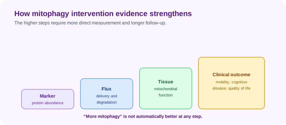
Mitophagy is a balance problem. Too little clearance can allow damaged organelles to accumulate, but excessive or mistimed clearance can remove mitochondria that remain useful, reduce energy supply or overload lysosomal systems. The desirable level likely depends on tissue, age, stress state, baseline damage and the capacity for biogenesis.
“Boosting mitophagy” is therefore incomplete without specifying which route, in which tissue, for how long and with what downstream effect. A marker can rise because initiation increased, because degradation stalled or because the cell is remodeling in response to stress. A decrease can mean less damage, impaired signaling or successful completion followed by recovery.
Safety also matters. A compound that changes mitochondrial turnover may interact with exercise adaptation, immune activation, medications or disease biology. Short-term tolerability cannot establish long-term safety, and a favorable biomarker cannot override symptoms or clinically abnormal tests.
Timing may be as important as magnitude. A short pulse during recovery from exercise or injury could have a different meaning from persistent pathway activation in a chronically stressed cell. Human studies should therefore report when samples were collected, whether the intervention changed mitochondrial content, and whether any functional benefit remained after the exposure ended.
Evidence verdict and translational gaps
Mitophagy is mechanistically central and clinically relevant, especially in disorders where mitochondrial damage and quality control are implicated. Human genetics, tissue studies and randomized intervention trials provide meaningful pieces of evidence. Yet those pieces do not currently support a general claim that a supplement, fasting protocol or drug slows human aging by increasing mitophagy.
The next generation of studies needs better human flux measures, tissue-aware sampling, adequate follow-up, preregistered primary endpoints and independent replication. Trials should distinguish pathway engagement from functional benefit and should report route, dose studied, tissue, comparator, adverse events and conflicts of interest clearly.
A practical evidence sequence is:
- identify the damaged mitochondrial process and target tissue;
- show that the intervention reaches the compartment and changes the intended pathway;
- demonstrate delivery and degradation rather than only static marker abundance;
- connect the pathway change to a prespecified physiological or patient-important outcome;
- test durability, safety and generalizability in adequately powered humans.
That standard keeps mitophagy in its proper place: an important biological mechanism whose therapeutic potential is real enough to study, but whose human longevity implications remain unproven.