What autophagy is
Autophagy is a family of lysosome-mediated pathways that remove and recycle intracellular material. The cargo can include proteins, lipid structures, damaged organelles, aggregates and other components. Recovered building blocks can support metabolism, adaptation, differentiation and survival when the cell is under stress.
The process is selective as well as bulk. A cell may increase general recycling during nutrient shortage while targeting a particular organelle through selective autophagy. Autophagy also participates in normal development and immune defense, so its function cannot be reduced to “clearing aging debris.”
Macroautophagy, microautophagy and chaperone-mediated autophagy
Macroautophagy encloses cargo in a double-membrane autophagosome before fusion with a lysosome. Microautophagy involves lysosomal or late-endosomal membrane uptake. Chaperone-mediated autophagy transports selected soluble proteins across the lysosomal membrane using chaperone and receptor machinery. These routes overlap in purpose but are not interchangeable measurements.
The foundational Nature Aging review of autophagy in healthy aging and disease describes this functional diversity and the continuing uncertainty about how autophagy, aging and disease interact. That uncertainty is a reason to define the pathway precisely, not a reason to discard it.
Autophagosome formation and lysosomal degradation
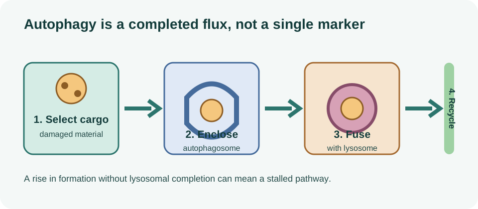
Macroautophagy begins with initiation and nucleation signals that organize a growing isolation membrane. The membrane expands around selected cargo, closes into an autophagosome and travels through the cell. Fusion with a lysosome exposes the cargo to enzymes that degrade it, after which metabolites can be returned to the cytosol.
Energy state, amino acids, growth factors, organelle contacts, membrane supply and stress signals all influence the pathway. ULK complexes, class III phosphatidylinositol 3-kinase complexes, ATG proteins, LC3-family conjugation and cargo receptors are parts of a coordinated system rather than independent switches.
A defect can occur at any step. More autophagosomes may mean increased initiation, reduced fusion, impaired lysosomal acidification or a stalled pathway. Conversely, fewer autophagosomes may reflect lower cargo burden or efficient completion. The same image can therefore support different interpretations depending on the assay and time point.
| Step | What researchers may observe | What it does not establish alone |
|---|---|---|
| Initiation | ULK or upstream signaling changes. | Successful cargo degradation. |
| Cargo capture | LC3, receptor or ubiquitin-associated structures. | Complete flux through the lysosome. |
| Fusion | Autophagosome–lysosome colocalization. | Efficient enzymatic breakdown. |
| Recycling | Turnover, isotope or metabolite recovery. | Improved healthspan in a person. |
Autophagic flux versus static markers
Autophagic flux describes movement through the pathway over time. Researchers may compare formation and clearance with lysosomal inhibitors, tandem fluorescent reporters, turnover labeling, microscopy, proteomics or other approaches. Each method has assumptions, and no single marker provides a complete readout in every tissue.
LC3-II accumulation can indicate more autophagosome membrane, but it can rise because formation increased or because degradation slowed. p62/SQSTM1 abundance can fall with degradation or change through transcription and stress responses. Lysosome number, acidity and protease activity add context but still do not automatically demonstrate beneficial recycling.
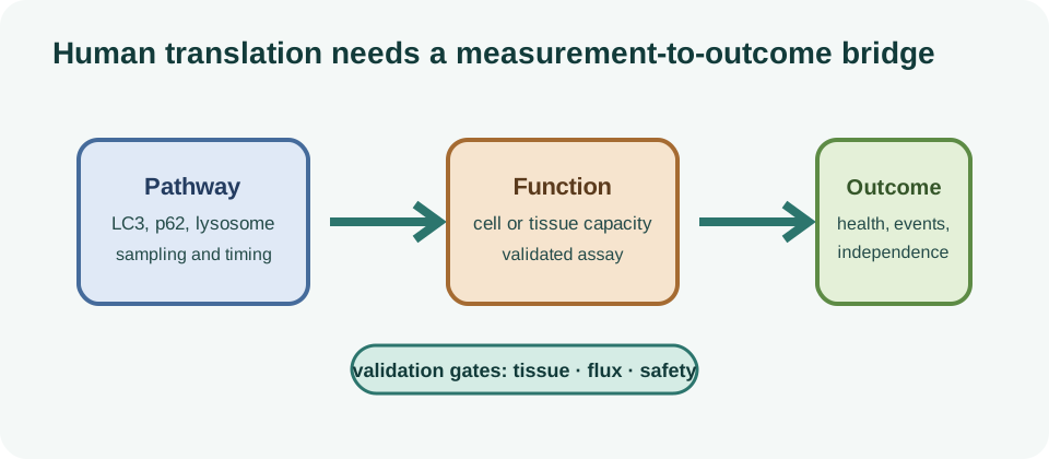
Human studies are especially constrained. Tissue biopsies sample a small compartment, blood measurements integrate organs and many assays measure expression rather than completed turnover. A reliable human endpoint would need validated sampling, timing, normalization and a demonstrated relationship with a clinically relevant outcome.
Why autophagy matters for proteostasis and organelle quality
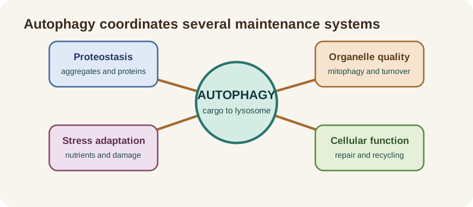
Autophagy complements the proteostasis network by clearing aggregates and damaged structures that chaperones or the proteasome cannot handle alone. It also intersects with mitochondria, endoplasmic-reticulum stress, peroxisomes, lipid droplets and immune signaling. The final result depends on the capacity of these systems to cooperate.
Selective mitochondrial autophagy, or mitophagy, is one important branch. It has its own cargo receptors, damage sensors, dynamics and lysosomal requirements. General autophagy markers should not be presented as direct measurements of mitophagy, and a mitophagy marker should not be treated as a complete readout of cellular autophagy.
Proteostasis failure can be both a cause and a consequence of aging-related dysfunction. Autophagy may remove harmful material, but persistent stress can also overload the pathway. The systems-level proteostasis and aging page provides that broader context, including why clearance and repair must be interpreted together.
How autophagy changes with age
Age-associated changes can affect autophagosome formation, cargo recognition, lysosomal acidity, membrane trafficking, transcriptional control and recycling. The pattern varies by tissue, cell type, sex, disease state, activity, nutrition and experimental model. A result in an old mouse liver should not be assumed to describe a human neuron or skeletal muscle.
The Nature Aging review notes that compromised autophagy is implicated in age-related pathology, while also emphasizing complex relationships rather than a single universal direction. Some age-related states may show reduced clearance; others may show stress-induced activation, compensation or dysregulation.
Age also changes the consequences of intervention. A young cell with abundant lysosomal reserve may respond differently from a senescent or diseased cell. Increasing initiation without fixing downstream degradation could increase cargo congestion. A modest pathway response that restores a specific tissue function could be more meaningful than a large marker change without benefit.
What model organisms can show about lifespan
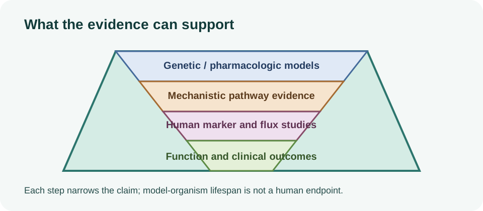
Genetic and pharmacological studies in yeast, worms, flies and mice provide some of the strongest causal evidence connecting autophagy-related pathways with lifespan or stress resistance. Loss-of-function experiments can show that an intervention’s effect depends on autophagy genes, while gain-of-function experiments can test whether pathway changes are sufficient in a model.
These experiments are valuable because they can manipulate tissues, timing and genes in ways that are difficult in humans. They also reveal that the result may depend on neuronal, intestinal, muscle, liver or immune-cell autophagy. A lifespan effect in a short-lived organism is a mechanistic signal, not a direct human clinical result.
The 2026 Nature Aging report on hypoxia-induced autophagic degradation and mammalian lifespan represents active frontier research, but it remains preclinical. It can motivate a human hypothesis while leaving open whether the same stimulus, tissue pathway and trade-offs operate in people.
Even within animals, “autophagy extends lifespan” is too broad. Some interventions may change autophagy as one part of a larger stress-response program. Genetic background, diet, pathogen status, housing and dose can alter the observed effect. Human translation requires independent trials with outcomes that matter to patients.
mTOR, AMPK and nutrient sensing
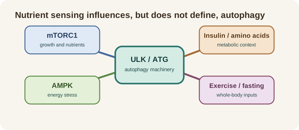
mTORC1 generally coordinates growth and nutrient availability, while AMPK responds to cellular energy stress. Their interaction with ULK signaling helps explain why starvation, exercise and some compounds can influence autophagy-related pathways. The pathway is integrated with insulin signaling, amino-acid sensing, mitochondrial metabolism and transcriptional programs.
The 2026 review of dietary restriction in aging and longevity places autophagy within a broader nutrient-sensing network. Dietary restriction can affect energy balance, substrate use, inflammation, hormones, circadian biology and behavior at the same time. A human outcome cannot be attributed to autophagy merely because an upstream signal changed.
Rapamycin and related mTOR-directed strategies are also not interchangeable with fasting or a general autophagy supplement. The mTOR and aging page covers the distinction between pathway modulation, dose studied, adverse effects and clinical endpoints. Mechanistic elegance does not remove the need for human safety and efficacy data.
Mitophagy is selective autophagy, not all autophagy
Mitophagy targets mitochondria for autophagic processing and depends on damage sensing, cargo selection, mitochondrial dynamics, trafficking and lysosomal completion. PINK1/Parkin is one important pathway, but receptor-mediated and other routes also operate in mammalian cells. The current Nature Reviews Molecular Cell Biology mitophagy review emphasizes this pathway diversity.
That distinction matters for interventions. A fasting-related change in a general autophagy marker may not mean damaged mitochondria were selectively removed. A rise in a mitophagy-related protein may not prove completed turnover. The experimental question must match the claim.
See the dedicated mitophagy evidence page for flux, tissue and Urolithin A context. The goal is a connected evidence map, not a collection of identical promises about “cellular cleanup.”
Spermidine and Urolithin A as study examples
Spermidine is an endogenous polyamine associated with autophagy-related biology and has been studied in observational and intervention research. Observational relationships can be confounded by diet, health status and behavior; a supplement trial must be judged by its prespecified outcomes, dose studied and duration. The spermidine evidence page follows those distinctions.
Urolithin A is a gut-microbe-derived postbiotic studied primarily in relation to mitochondrial quality and mitophagy. Human randomized trials have reported selected muscle, endurance and mitochondrial-related results, but they do not establish that general autophagy was increased throughout the body or that lifespan was extended. The Urolithin A page records its endpoint-specific evidence.
| Intervention question | Evidence needed | Common overclaim |
|---|---|---|
| Spermidine | defined exposure, human outcome and relevant pathway measure. | dietary association proves lifespan extension. |
| Urolithin A | mitochondrial or muscle endpoint in the studied population. | muscle signal proves whole-body autophagy. |
| Fasting | validated human flux measurement with controlled timing. | a universal hour threshold applies to everyone. |
What human intervention studies can measure
Human studies can measure body composition, metabolic markers, exercise performance, cognition, symptoms, inflammatory markers, tissue transcripts, proteins, mitochondrial function and selected autophagy-related signals. A biopsy can add local evidence, but it is invasive and not necessarily representative of every organ. Blood is easier to collect but often less specific.
The strongest design pairs a pathway measure with a prespecified functional or clinical endpoint and follows participants long enough to assess durability. It reports randomization, comparator, adherence, missing data, adverse events, multiple-testing decisions, tissue and sampling time. A small biomarker pilot can be useful without being a clinical efficacy trial.
Human intervention results can also be null. A null primary endpoint does not mean the pathway is irrelevant; it may mean the dose, tissue, timing or target was wrong. It does mean a broad benefit claim is not supported by that study. The hierarchy should remain clear: molecular signal, physiological response, function, disease outcome and longevity are distinct levels.
Why “fasting for X hours turns on autophagy” is overclaimed
Fasting changes nutrient availability and can influence insulin, glucagon, AMPK, mTOR, substrate use and other signals that interact with autophagy. But the timing and magnitude depend on the tissue, preceding diet, activity, body composition, sleep, illness, medication, sex and metabolic state. A precise universal hour threshold is not established by the available human evidence.
Researchers may observe fasting-related changes in blood metabolites or in animal tissues, but those findings do not prove that a person’s brain, liver, muscle and immune cells all entered the same autophagic state. Nor do they show that a marker change completed lysosomal flux or improved health.
Fasting can also carry risks for people with diabetes, eating-disorder history, pregnancy, frailty, medication-sensitive conditions or inadequate nutritional reserve. A general article should not turn an uncertain mechanistic timing claim into a personalized fasting schedule.
Potential risks of too little or dysregulated autophagy
Insufficient autophagy can impair clearance of damaged proteins and organelles, contribute to stress sensitivity and interact with age-related disease biology. But indiscriminate activation can also be harmful. Autophagy may support survival in some cancer contexts, participate in infection responses and influence tissue remodeling. Its effect is context-dependent rather than uniformly protective.
Interventions that manipulate mTOR, AMPK, lysosomes or nutrient intake may affect immune function, fertility, glucose control, muscle mass, medication exposure or disease progression. A pathway change should not be treated as inherently beneficial. Safety monitoring and clinical context remain necessary even when a compound is marketed as “natural.”
Excessive or stalled autophagy can also be confused with successful clearance. If lysosomal degradation is impaired, upstream markers may rise while cellular waste accumulates. Conversely, strong clearance in one tissue may deplete resources if replacement and metabolic support are inadequate.
For that reason, a sensible intervention study should define a stopping rule for adverse events and prespecify which biomarker, function measure and clinical outcome matter most. It should not assume that a favorable molecular signal offsets hypoglycemia, malnutrition, loss of lean mass, infection risk or a worsening disease marker. These are trial-design and clinical-monitoring questions, not arguments for or against autophagy in the abstract.
Evidence verdict and research gaps
Autophagy is indispensable biology and a credible target for research into proteostasis, organelle quality, stress adaptation and age-related disease. Model organisms provide causal evidence that some lifespan-extending interventions depend on autophagy-related pathways. That evidence does not establish a human “autophagy score,” a universal fasting threshold or a general anti-aging treatment.
Progress depends on validated human flux measures, tissue-aware sampling, longitudinal design, prespecified clinical endpoints and independent replication. Studies should report what was measured, what was not measured, which dose was studied and how safety was assessed. They should also distinguish general autophagy from mitophagy and separate biomarker movement from functional benefit.
- define the cargo, tissue and pathway before choosing a marker;
- measure flux or turnover rather than relying on static abundance alone;
- connect pathway engagement to a prespecified physiological or patient-important endpoint;
- test durability, safety and generalizability in humans;
- retain null and contradictory findings in the evidence record.