Seeing a tooth outline or a darker area through the gum does not automatically mean the gum has receded. Some people naturally have thin gum tissue over prominent roots, while true recession means the gum edge has moved away from its earlier position and more root surface is exposed. These can look similar in a mirror, but they are not the same finding and do not call for the same treatment.

What does it mean when you can see a tooth through the gum?
The visible part of a tooth is its crown. The root normally sits under the gum and is supported by the periodontal ligament and jawbone. Gum tissue covers and protects the supporting structures around each tooth. Its thickness and shape vary between people and between different areas of the same mouth. When tissue is thin and a root is prominent, the root’s outline or darker color may show through the gum even if the gum edge has not moved.
Periodontal phenotype describes features such as gum thickness, the band of keratinized tissue, and the shape of the supporting bone. A 2019 American Academy of Periodontology evidence review found that thin or narrow gingiva is associated with a greater prevalence or severity of recession at affected sites. That association does not mean every person with thin tissue has disease, or that healthy thin tissue must be thickened. The review found no evidence supporting routine conversion of healthy thin sites to a thicker phenotype when there is no recession or mucogingival defect.
By contrast, gingival recession describes the gum margin moving toward the root, away from the crown. This can expose a root surface that is normally covered. A person may notice a tooth looks longer, a notch or yellowish area near the gumline, a new space between teeth, or sensitivity to cold, sweets, brushing, or touch. Recession can occur with or without periodontitis, so the word “recession” alone does not identify its cause.
Thin tissue versus actual gum recession
Clues can help you decide whether to book an assessment, but they cannot replace one. A long-standing, similar appearance on both sides with a stable gum edge may fit naturally thin tissue. A newly exposed root, a gumline that is moving, or a tooth that seems to be getting longer deserves evaluation. Bleeding or swelling can indicate inflammation, but the absence of bleeding does not rule out a problem.
Researchers have also studied whether a periodontal probe showing through the gum can classify thin versus thick tissue. A 2024 systematic review and meta-analysis found that this transparency method has limited diagnostic accuracy, with results depending on the measurement threshold and location. It is one clinical clue, not a standalone test. The dentist may combine visual examination with probing measurements, medical and dental history, tooth position, and radiographs when indicated.
Possible reasons a gumline changes
There is no single cause for every area of recession. Periodontitis can damage the attachment and supporting bone around teeth. Repeated traumatic brushing can injure tissue, while smoking or tobacco use raises periodontal risk and can affect healing. Thin tissue, a prominent or poorly positioned tooth, previous trauma, and oral piercings may also matter. Orthodontic tooth movement is another factor to discuss in context: movement within the bony housing may be appropriate, but moving a tooth outward through thin or limited bone can raise concern for recession.
Plaque-related inflammation is relevant, but blaming every receding gumline on “poor hygiene” is inaccurate. Some people develop recession despite careful daily cleaning because anatomy, tissue thickness, tooth position, brushing trauma, past treatment, or other local factors are involved. Conversely, an apparent increase in tooth length can have causes other than recession, so a clinician should check rather than assume.
- Periodontal inflammation or periodontitis: assessment considers pocket depths, attachment levels, bleeding, and supporting bone.
- Mechanical factors: forceful scrubbing, injury, or pressure from a piercing may affect a local area.
- Anatomy and tooth position: thin tissue or a root prominent in the jaw can make an area more vulnerable or visible.
- Tobacco exposure or treatment history: these details help a clinician understand risk and healing.
If you are already receiving orthodontic treatment, tell your orthodontist promptly about a new change in the gumline. Do not stop or alter prescribed appliances on your own; the treating clinician can assess tooth movement, tissue health, and whether the plan needs adjustment. For general questions about alignment and gum support, see our guide to orthodontic specialists and general dentists.
How a dentist checks the gumline
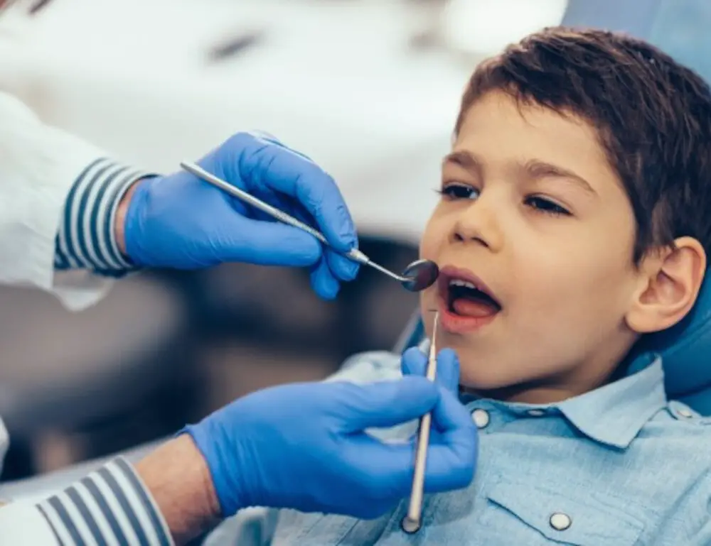
An evaluation usually starts with when you first noticed the change, whether it is progressing, and whether you have sensitivity, bleeding, swelling, pain, or a recent injury. The clinician inspects the gum margin and nearby teeth, checks for plaque and inflammation, and measures around the teeth with a periodontal probe. Recession and pocket depths are recorded separately because a visible root does not tell the whole story about attachment or bone support.
Depending on the findings and your history, dental radiographs may help assess supporting bone or other conditions. A radiograph is not a direct measurement of soft-tissue thickness, and a single photo cannot show the full periodontal diagnosis. Probe transparency is also imperfect. The goal is to determine whether tissue is simply thin and stable, whether the margin has receded, whether periodontal disease is present, or whether another issue such as root-surface wear or decay explains the appearance.
Ask the dentist to show you where the gum margin is now and whether there are previous measurements or images for comparison. Useful questions include: Is this true recession? Is there attachment or bone loss? Is the area stable? What is causing sensitivity, if present? Which changes should prompt an earlier follow-up? If you have a history of gum disease, you can also review our overview of healthy gums, gingivitis, and periodontal disease.
Treatment depends on what the examination finds
Stable, healthy thin tissue
If the gum margin is stable, there is no active periodontal disease, and the root is simply visible through naturally thin tissue, treatment may not be needed. Monitoring during routine dental visits and gentle plaque control may be enough. There is no established need to graft or otherwise thicken every healthy thin area solely because an outline can be seen. A clinician may recommend closer monitoring if the site has risk factors or changes over time.
Inflammation or periodontitis
When gum inflammation or periodontitis is present, treatment targets that disease and its causes. Professional cleaning, home-care coaching, and—when diagnosed periodontitis warrants it—scaling and root planing can be part of care. Our overview of periodontal disease and tooth support explains why gum health and supporting bone matter over time. The American Dental Association describes scaling and root planing as a nonsurgical treatment for periodontitis. It is not a procedure that makes receded gum grow back simply because a root is visible. Antibiotics or antimicrobial rinses may have selected uses under professional direction, but they are not generic treatments to restore gum coverage.
Exposed-root sensitivity
For sensitivity, a dentist may suggest a desensitizing toothpaste, fluoride varnish, or another treatment based on the cause. Root-surface decay, a crack, wear, or a problem with the nerve may need different care. Use a soft-bristled toothbrush and light pressure; aim to clean along the gumline without forceful horizontal scrubbing. Continue daily interdental cleaning as advised rather than avoiding a sensitive area altogether. Our guide to flossing and bleeding gums explains why technique and persistent symptoms matter.
Selected recession defects
For some recession defects, a periodontist may discuss periodontal plastic surgery or a gum graft to increase tissue thickness, cover some exposed root, reduce sensitivity, or help protect the site. Whether coverage is achievable and how much is realistic depend on the defect, neighboring tissue, bone support, and treatment goals. A graft does not guarantee complete or permanent root coverage, and it is not needed for every visible root.
Pocket-reduction surgery is used in selected periodontal cases to improve access and reduce deep pockets; it is not the standard cosmetic technique for covering an exposed root. Removing additional tissue through gum contouring would generally not address thin or receded gum and could make the appearance worse. Whitening and veneers change tooth color or shape, not gum thickness or root coverage. If appearance is your main concern, discuss the diagnosis and trade-offs with a dentist before considering a cosmetic procedure.
What you can do while waiting for an appointment
- Brush twice daily with a soft-bristled brush and gentle pressure, using fluoride toothpaste.
- Clean between teeth daily with a method that fits your mouth; ask a dental professional for technique help if flossing causes persistent bleeding or pain.
- Avoid tobacco and tell your dental team about tobacco use, piercings, orthodontic treatment, and recent changes.
- Note when you first noticed the outline, whether it is changing, and what triggers sensitivity; bring earlier photographs only as context.
These steps support oral hygiene; they do not diagnose or reverse recession. Do not stop prescribed periodontal or orthodontic care because a page or product promises to “regrow” gum tissue. If symptoms continue, ask for a clinical assessment and an explanation of the measurements and options. For a broader view of gum symptoms and prevention, see why gums may bleed during brushing and common causes of bleeding gums.
When to arrange an evaluation
Book a dental or periodontal evaluation if the gumline is moving, a root has become newly exposed, sensitivity is increasing, or you notice persistent bleeding or swelling. Arrange prompt care for pus, a loose tooth, significant pain, or a sudden change after trauma. If a new or worsening gumline change appears during orthodontic treatment, contact the treating orthodontic team. These signs do not prove a specific diagnosis, but they are reasons to have the area examined.
Stable appearance over a long period without symptoms may be less urgent, but mention it at a routine visit so the clinician can record the baseline. A baseline makes it easier to distinguish a long-standing thin phenotype from a new change at a later appointment.
Frequently asked questions
Can gums be transparent without receding?
Yes. Naturally thin tissue over a prominent root may make an outline or darker area visible even when the gum margin has not moved. An examination is needed to confirm that the tissue is healthy and stable.
Does seeing a root mean I have gum disease?
No. A visible root can occur with recession, but recession can have several causes and is not itself enough to diagnose periodontitis. A dentist checks the gum margin, pocket depths, attachment, inflammation, and supporting bone as appropriate.
Can receded gums grow back on their own?
Lost gum coverage does not normally return on its own. Some selected defects can be treated with grafting, but the expected coverage varies. Thin tissue without recession may need no treatment at all.
Will a deep cleaning cover an exposed root?
No. Scaling and root planing treats diagnosed periodontitis when indicated; it does not regrow gum tissue. The clinician can explain whether periodontal treatment, sensitivity care, monitoring, grafting, or another option fits the findings.
Can braces or aligners fix gum recession?
Orthodontics is not a general recession treatment. Tooth movement may be appropriate in some cases, but movement through thin or limited supporting bone may increase risk. A plan should consider gum health, tooth position, and bone support.