Scope note: This study explainer is not an exercise prescription. The trial tested specific supervised programs in a selected group; individual activity choices should account for health, function, experience and clinical advice where relevant.
What question did the LISA brain-clock analysis ask?
Resistance training is usually evaluated through outcomes such as strength, mobility, physical function or cognition. Neuroimaging can add another layer by measuring brain structure or function, but a change in an image is not automatically a change in memory, independence or disease risk. Brain-age models compress patterns across a scan into an estimate that is compared with chronological age. This secondary analysis asked whether those model-based estimates changed across the LISA trial’s training groups.
The distinction matters because the article’s headline word “clock” can sound more definitive than the underlying instrument. Here, the clock used resting-state functional MRI connectivity features. It did not count years of life, diagnose brain disease, or measure a person’s complete aging process. The study’s score is best treated as a research biomarker that may help generate hypotheses about how training relates to distributed brain networks.
The 2026 GeroScience paper reanalyzed imaging from LISA rather than reporting a new, stand-alone randomization. Its experimental foundation is useful, but the analyzed imaging subset and model validity still need separate scrutiny.
Who took part, and what were the training groups?
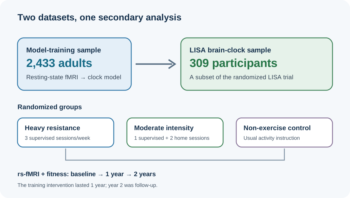
The brain-clock analysis included 309 adults aged 62–70 from LISA, a randomized, supervised one-year training trial in Denmark. Participants were assigned to heavy resistance training, moderate-intensity training, or a non-exercise control. The analysis paper describes a healthy older sample; it should not be assumed to represent people with substantial illness, neurological conditions or musculoskeletal limitations.
The heavy group attended three supervised sessions per week at a training center. The moderate-intensity group attended one supervised session and completed two home-based sessions each week. Both exercise programs combined resistance work with functional training intended to improve strength, endurance and balance. The control group was asked to maintain usual activity and avoid more than about one hour of strenuous activity per week. These were the trial’s defined programs, not universal recommendations.
The imaging subset is distinct from both the model-training sample and the full LISA enrollment. MRI and fitness were assessed at baseline, year one and year two; the program lasted one year, making year two follow-up rather than a second year of the same supervised training.
How was the brain-age score constructed?
Researchers trained LightGBM models on 6,670 resting-state connectivity features from 2,433 external adults, then applied them to LISA. The training set averaged about 62 years of age and spanned roughly 31–89. The pipeline used cross-validation and an age-bias correction for the predicted brain-age gap.
For each scan, predicted brain age was compared with chronological age to form a brain-age gap (BAG). A lower score over time can be described as a more favorable model estimate under this pipeline. It cannot by itself establish that neural tissue has reversed its age, or that the score maps onto a particular number of years of preserved cognition or life.
The model’s reported mean absolute error was about 8.65 years in its training and validation procedures. That figure is not a confidence interval for the group change, and it does not mean the study’s within-person analyses are automatically meaningless. It does underscore that the model is noisy as an individual age estimator. The authors used it to compare patterns statistically across a group; it is not a clinical instrument for telling a person that their brain is a precise number of years younger or older.
What brain-age-gap changes were reported?
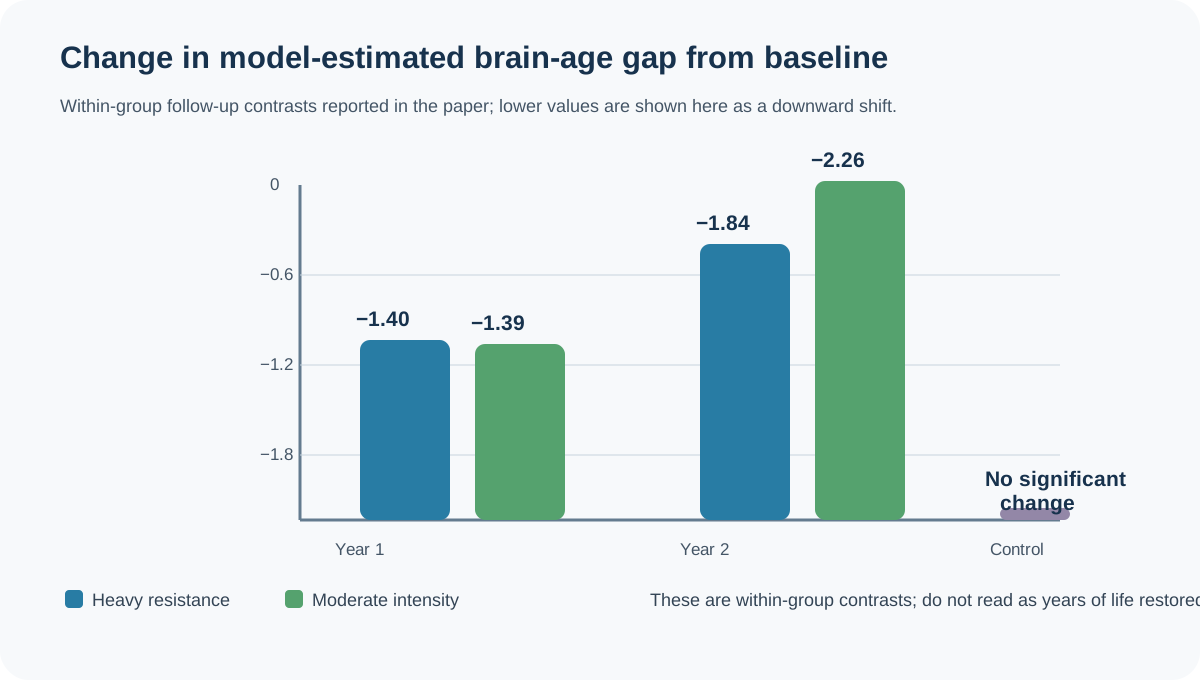
The paper reported lower brain-age-gap estimates in both exercise groups at follow-up. In the heavy-resistance group, the reduction relative to baseline was 1.40 years at the one-year assessment and 1.84 years at year two. In the moderate-intensity group, the corresponding reductions were 1.39 and 2.26 years. The non-exercise group did not show a statistically significant within-group change. The reported group-by-time model also found an interaction, and the study adjusted the set of group/time comparisons using the Benjamini–Hochberg false-discovery-rate procedure.
| Assigned group | Baseline to year 1 | Baseline to year 2 | How to read it |
|---|---|---|---|
| Heavy resistance | −1.40 years | −1.84 years | Lower model-estimated BAG within group |
| Moderate intensity | −1.39 years | −2.26 years | Lower model-estimated BAG within group |
| Non-exercise control | No significant within-group change reported | Not evidence that the control group’s brain became older | |
These are changes from baseline within each assigned group. They are not direct evidence that the moderate program outperformed the heavy program, and the 2.26-year value is not a guaranteed effect for an individual. The ranges summarized in the abstract (about 1.4–2.3 years) combine the reported significant results. They should not be rewritten as “resistance exercise turned back brain age by two years.”
What did the connectivity analysis add?
The paper also tested local functional-connectivity changes from baseline to the end of the one-year intervention. Compared with the non-exercise control, the heavy-training group showed a significant cluster, primarily in prefrontal regions with additional motor-cortex and superior-parietal involvement. The reported cluster contained 8,517 voxels and had a cluster-level false-discovery-rate value below 0.001. The moderate group did not differ significantly from control in this analysis, and the heavy and moderate groups did not significantly differ from one another.
This localized result and the brain-clock result are related but distinct analyses. The clock summarized connectivity across thousands of features; the voxel-level analysis mapped where a group contrast appeared in the scan. The regional finding does not show that executive function improved, even though the prefrontal cortex contributes to functions such as planning and control. Cognitive tests and clinical events are separate endpoints.
The researchers also explored brain clocks limited to individual resting-state networks, including default-mode, motor, somatosensory and cerebellar networks. Those network-specific interaction tests were not statistically significant. That weakens any claim that one named network was clearly responsible for the whole-brain clock result. It also illustrates how conclusions can depend on whether a model is built from distributed connectivity or a selected network subset.
What can randomization establish here?
Random assignment is a meaningful strength. If follow-up and analysis preserve the randomized comparison, it helps reduce the baseline confounding that complicates observational exercise studies. This makes the trial more informative about whether offering these specific training programs changed the measured imaging-derived outcomes in the analyzed participants.
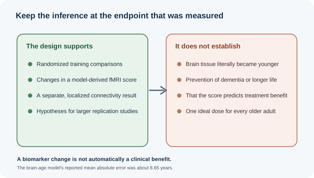
Randomization does not make every later interpretation causal. This paper is a secondary brain-clock analysis of a subset with the required imaging data. The clock itself was trained in an external dataset; a randomized intervention cannot validate the score as a surrogate for cognition or dementia. Statistical changes in an intermediate measure do not prove that participants experienced a clinical benefit, and a biomarker can move without predicting a meaningful outcome.
Within-group changes are not treatment-effect estimates. The paper used group-by-time models and multiplicity-adjusted comparisons; those direct contrasts and their uncertainty matter more than comparing separate “significant” values. The result identifies no optimal dose and does not show heavy resistance is superior.
What are the key limits and how does this differ from AGUEDA?
The analysis was limited to a 309-person imaging subset of healthy older adults from a high-income European setting. The findings may not transfer to people with major comorbidities, neurological disease or different social and health contexts. MRI-based research also depends on scan quality, preprocessing choices, motion control and how the model was trained. The authors report a model MAE of about 8.65 years, which cautions against individual-level interpretation.
The analysis also found no significant clock effect in individual network-restricted models and only a modest relationship between BAG change and leg-strength change overall (Spearman r = −0.12, p = 0.036). The within-group association appeared in the moderate group, not the heavy or control group; it does not show that strength gain mediated the brain-score change.
Article 189’s AGUEDA work addresses a different randomized resistance-exercise analysis, focused on Alzheimer-related blood, amyloid and MRI signatures. The LISA paper here focuses on resting-state functional connectivity and a machine-learning brain-age-gap estimate. The studies cannot be merged into a single claim that resistance exercise prevents Alzheimer’s disease. Brain-age clocks, Alzheimer-related signatures, strength, cognition and dementia diagnoses are not interchangeable outcomes.
What would strengthen the evidence next?
A useful next step is independent replication of the brain-clock result in a larger randomized sample with prespecified imaging outcomes, transparent retention data and accessible analysis code. Researchers should report direct group-by-time contrasts, confidence intervals, sensitivity to motion and preprocessing, and whether a brain-age score adds information beyond standard measures of function and cognition.
Clinical follow-up should test whether score changes track changes people can notice or value: cognition, daily function, falls, disability or diagnosed disease. Surrogate validation requires more than a correlation in one trial; it needs evidence across studies that intervention-related changes in the marker reliably predict intervention-related changes in clinical outcomes. Longer follow-up and diverse participants would help address generalizability and durability.
A reader can reasonably take away that one randomized-trial secondary analysis found lower model-estimated BAG after two defined resistance-training programs, with an additional heavy-versus-control connectivity contrast. Readers should not infer that a brain clock measured literal age reversal, that a particular program prevents dementia, or that every older adult should follow the trial schedule. The study is a research signal, not a clinical decision rule.
Frequently asked questions
References
Internal reading
For related topics, see brain aging, sarcopenia and muscle aging, biological-age testing, the Systems hub, and the Interventions hub.