What proteostasis means
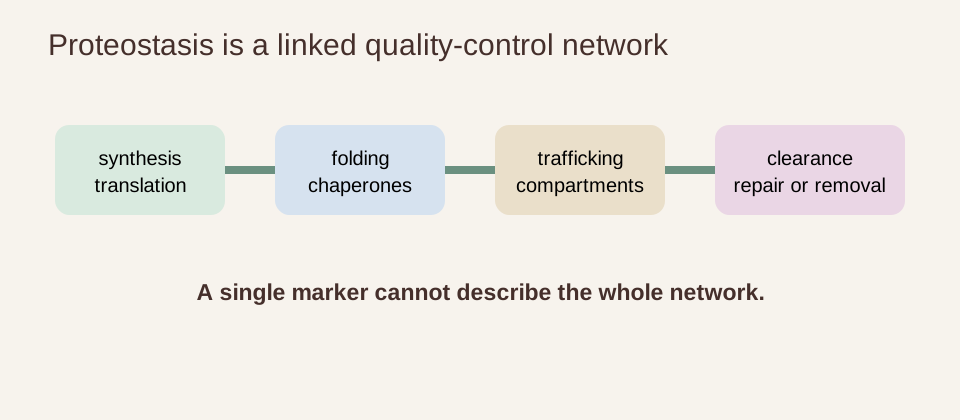
Proteostasis is the network that keeps a cell’s proteins functional over time. It begins with transcription and translation, continues through folding and trafficking, and ends with repair, recycling or degradation when a protein is damaged or no longer needed. Chaperones, the ubiquitin–proteasome system, autophagy, lysosomes and organelle stress responses all contribute.
The network is dynamic rather than static. Cells alter protein synthesis during fasting, infection, heat, oxidative stress and nutrient change. A temporary stress response can be protective, while chronic stress can exhaust repair and clearance capacity. Greater clearance is not automatically beneficial: indiscriminate removal can also eliminate essential proteins or healthy organelles, so the cargo and tissue context matter.
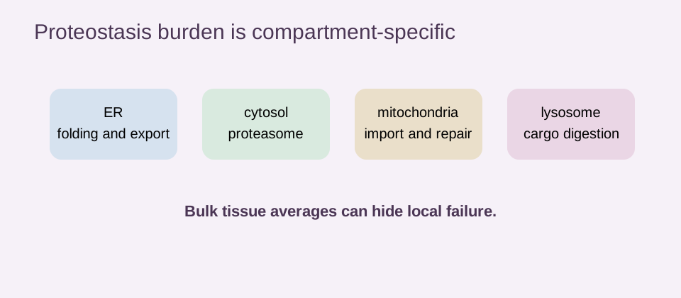
Loss of proteostasis is therefore a systems concept. Protein abundance, aggregation, turnover and activity are distinct measurements. A protein can be abundant but inactive, reduced because it was properly cleared, or apparently normal in bulk while damaged in a long-lived cell.
Protein synthesis, folding, trafficking and degradation form one network
Ribosomes make polypeptide chains that must fold into precise three-dimensional structures. Chaperones help new or stressed proteins reach functional conformations. The endoplasmic reticulum folds secreted and membrane proteins, adds modifications and sends correctly processed cargo onward. Trafficking errors can cause proteins to accumulate in the wrong compartment.
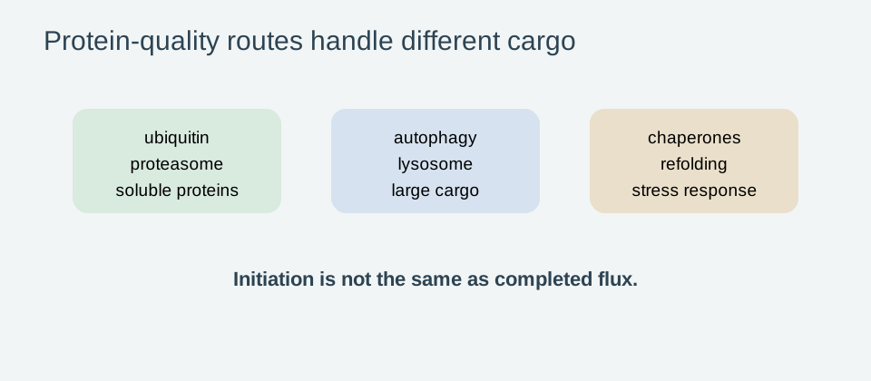
Misfolded or damaged proteins may be tagged with ubiquitin and directed to the proteasome. Larger assemblies, damaged organelles and long-lived aggregate-prone material can be handled through autophagy and lysosomal degradation. These routes overlap, compensate for one another and compete for energy and raw materials.
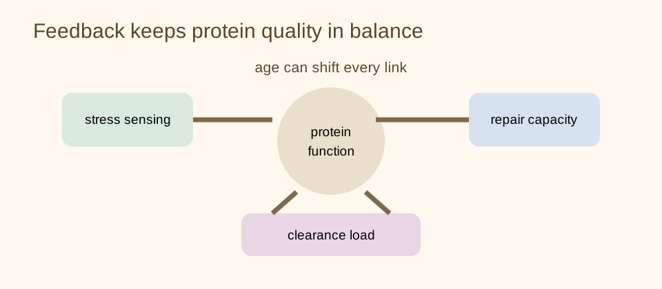
Age-related decline can occur at several points: translation may become less accurate, chaperone capacity may fall, proteasome activity may change, lysosomal clearance may slow, or stress signaling may become poorly coordinated. A study measuring only one component cannot describe the entire network.
| Measure | What it captures | What it cannot establish alone |
|---|---|---|
| Protein abundance | amount detected in a sample | correct folding, activity or turnover |
| Aggregate burden | insoluble or misassembled material | the sole cause of aging |
| Proteasome activity | one degradation route | whole-cell protein quality |
| Autophagy marker | one stage of a dynamic pathway | completed lysosomal flux |
Chaperones and heat-shock responses
Chaperones bind unstable proteins, prevent inappropriate interactions and help refold or route damaged proteins. Heat-shock factors coordinate a protective transcriptional response when proteotoxic stress rises. These systems can preserve function during acute stress and are part of the reason mild stress is studied in longevity biology.
Age can weaken the ability to induce or resolve a heat-shock response. Chronic inflammation, oxidative damage, altered translation and mitochondrial stress may increase the load placed on chaperones. Yet a permanently elevated stress response is not a simple health goal. It can consume energy, alter signaling and interact with immunity.
Small molecules that influence chaperones or heat-shock factors have shown promising effects in cells and animals. Human translation requires selectivity, tissue exposure and clinical outcomes. A compound that improves aggregation in a cultured cell is not established as a human anti-aging therapy.
Ubiquitin–proteasome, autophagy and lysosomal degradation
The ubiquitin–proteasome system removes many short-lived and soluble proteins. Autophagy delivers selected cargo, protein complexes and organelles to lysosomes. These systems are not interchangeable, and measuring a single upstream marker can be misleading. Increased autophagy initiation does not necessarily mean increased completed flux through lysosomal degradation.
Autophagy is frequently described as a universal longevity pathway. The more accurate view is context-dependent. The amount, timing and cargo of autophagy matter, as do nutrient status, tissue, age and disease. Excessive or poorly targeted degradation could be harmful, while insufficient clearance can permit toxic accumulation.
The autophagy and longevity page and mitophagy page distinguish pathway activation from demonstrated healthspan benefit. A human intervention must show that the intended process changed in the relevant tissue and that a meaningful clinical outcome improved.
Endoplasmic-reticulum stress and the unfolded-protein response
The endoplasmic reticulum, or ER, folds proteins destined for secretion or membranes. When unfolded proteins accumulate, the unfolded-protein response reduces some translation, increases chaperone capacity and promotes removal of irreparably damaged material. The response can restore balance, but persistent ER stress can trigger inflammation, dysfunction and cell death.
A 2025 review describes the ER proteostasis network as central to healthy aging across model systems and human tissue. This supports the importance of ER quality control as a research target. It does not identify one supplement, diet or drug as a proven way to restore ER proteostasis throughout the body.
ER stress is also linked with metabolic disease, neurodegeneration and immune signaling. A disease-specific therapy may reduce ER stress as part of treating that disease without slowing aging in healthy people. The claim must remain as narrow as the trial evidence.
Organelle-specific proteostasis and vulnerable neurons
Mitochondria, the ER, lysosomes, peroxisomes and the nucleus each face distinct protein-quality challenges. Mitochondrial proteostasis includes import, folding, protease activity and mitophagy. Lysosomal proteostasis depends on acidification, enzymes and cargo delivery. A global protein measurement may miss a compartment-specific failure.
Neurons are especially vulnerable because many are long-lived, highly polarized and difficult to replace. They must transport proteins over long distances and maintain synapses for decades. Age-related defects in clearance, trafficking or mitochondrial quality control can therefore accumulate. This helps explain why proteostasis is prominent in neurodegenerative-disease research.
Vulnerability does not mean protein aggregation is the sole cause of brain aging. Vascular injury, inflammation, synaptic change, metabolic stress and cell loss also matter. The brain-aging page keeps proteostasis within that broader network rather than treating one aggregate as a complete diagnosis.
Protein aggregation and amyloid accumulation with age
Aggregation occurs when proteins misfold, become insoluble or assemble into larger structures. Some aggregates are toxic, some are protective sequestration responses, and some may be inert bystanders. Their effects depend on composition, location, clearance and the cell’s ability to adapt.
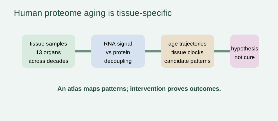
The 2025 Cell human atlas analyzed 516 samples spanning 13 tissues across five decades. It reported widespread transcriptome–proteome decoupling, proteostasis decline and amyloid accumulation, along with tissue-specific proteomic age trajectories. This is strong descriptive evidence that protein-level aging is observable in humans.
Descriptive evidence still has limits. The atlas does not show that amyloid accumulation is the sole driver of aging, nor that clearing it in a healthy person extends life. It identifies patterns and candidate mechanisms that require longitudinal and intervention studies.
The 2025 human multi-tissue proteome atlas
The atlas is important because it looks beyond blood and beyond one organ. Its tissue-specific analyses suggest that organs age at different rates and that protein abundance does not always track transcript abundance. That decoupling matters: measuring RNA alone can miss post-transcriptional, translational, folding and degradation changes.
The authors developed tissue-specific proteomic age clocks and reported an aging inflection around age 50 in their temporal analysis, with blood vessels among the tissues showing earlier susceptibility. These are model-derived trajectories, not a universal timetable for every individual. A clock or inflection point should not be used to diagnose a person’s proteostasis state.
The study also reported a plasma signature connected to tissue origins and candidate senoproteins, including GAS6. Candidate status is not proof of causality. The value of the atlas is that it maps hypotheses for validation, not that it supplies a ready-made anti-aging target.
The atlas is best read as a reference map: it shows where age-related protein changes are detectable and which questions deserve longitudinal testing. It does not tell a reader which supplement to take or which protein to remove. That restraint is important because the same observed protein pattern may be a cause, consequence or compensation for tissue stress, and future randomized studies may revise the interpretation outside this dataset in future evidence reviews.
Transcriptome–proteome decoupling and translation changes
Transcripts are instructions; proteins are the functional products that must be folded, modified, trafficked and removed. Their quantities can diverge because translation efficiency, degradation, localization and post-translational modification change. This is why transcriptomics alone cannot provide a complete proteostasis assessment.
A 2025 review of translational regulation in stress biology connects protein synthesis control with oxidative stress, nutrient sensing and age-related changes. Translation can be reduced to conserve resources during stress, while selective translation preserves stress-response proteins. With age, the balance may become less precise or slower to recover.
These mechanisms connect proteostasis with mitochondrial dysfunction and cellular senescence. They do not imply that activating one pathway always restores the network. The intervention, tissue, duration and downstream function still need to be demonstrated.
Preclinical strategies and why disease therapies are not automatically geroprotectors
Preclinical strategies include chemical chaperones, heat-shock modulation, proteasome tuning, unfolded-protein-response control, translation regulation, autophagy enhancement and targeted clearance of aggregates or damaged organelles. In cells and animals, some approaches improve stress resistance or disease phenotypes, and some extend lifespan under defined experimental conditions.
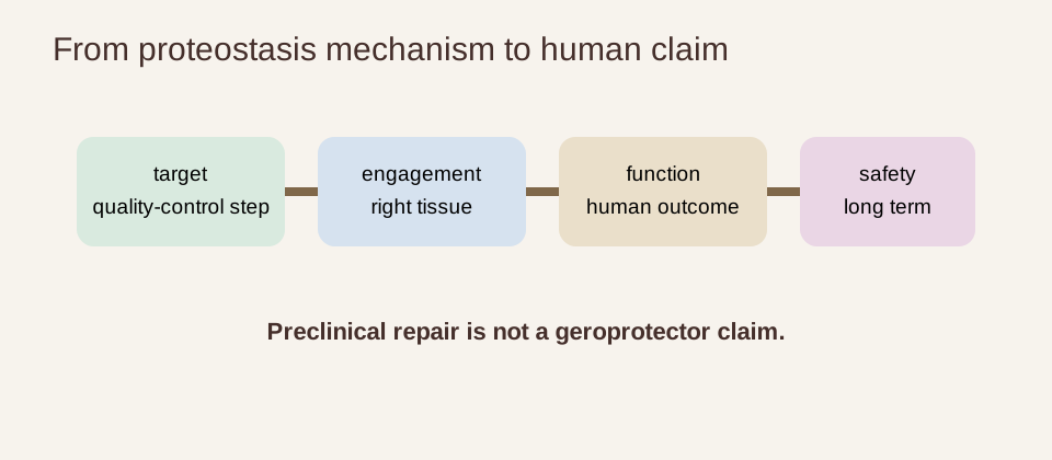
Translation requires more than a favorable model. A candidate must be selective, reach the relevant tissue, preserve essential proteins, avoid harmful immune or growth effects and produce a durable functional benefit. A neurodegenerative-disease treatment can reduce a disease biomarker without changing normal aging. A cancer therapy can alter proteostasis because tumor cells are unusually dependent on protein quality control, but that does not make it a healthy-aging therapy.
Human trials should distinguish target engagement, disease treatment, symptom improvement and geroscience outcomes. They should report function, disability, cognition, falls, disease events or survival where appropriate, not only aggregate burden or a proteomic score. The biology of aging hub keeps these evidence layers connected but separate. The National Institute on Aging biology-of-aging program also illustrates why mechanisms must ultimately be connected to measurable human outcomes.
Trial interpretation should also account for baseline disease and treatment intensity. A patient with a defined protein-misfolding disorder may respond differently from a healthy older adult with ordinary age-related changes. The relevant comparator may be standard care rather than placebo, and a short reduction in a disease marker may not predict durability. Proteostasis is a plausible therapeutic domain, but the indication and outcome must remain as specific as the study design.
Evidence verdict and translational requirements
Proteostasis is a central aging network that spans synthesis, folding, trafficking, chaperones, the proteasome, autophagy, lysosomes and organelle stress responses. Age-related decline is biologically plausible and increasingly visible in human tissue. The 2025 multi-tissue Cell atlas provides important descriptive evidence of proteostasis decline, amyloid accumulation and transcriptome–proteome decoupling across 13 tissues.
That evidence does not prove that aggregates are the sole cause of aging or that directly activating autophagy, chaperones or clearance will extend human life. Most direct restoration strategies remain preclinical. Disease therapies may act on proteostasis while treating a disease, but their effects should not be generalized to healthy longevity.
The responsible conclusion is human proteostasis decline is real, while direct proteostasis rejuvenation remains experimental. A convincing geroscience intervention would demonstrate tissue-specific target engagement, improved protein quality or flux, preserved organelle function, meaningful functional outcomes and acceptable long-term safety. Protein abundance, aggregate burden or a proteomic age score alone cannot carry the claim.
- separate protein abundance, aggregation and turnover;
- identify the compartment and tissue being measured;
- treat autophagy and stress markers as dynamic pathway evidence;
- keep human descriptive atlases separate from lifespan interventions in animals;
- require functional and safety outcomes before using rejuvenation language.