What the July 2026 Nature Medicine review argues
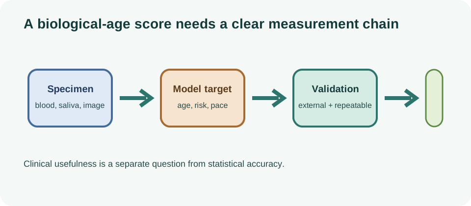
The review presents biological aging clocks as statistical tools that estimate age-related features in an individual, organ, tissue or cell. Their potential uses include risk stratification, prevention research and monitoring interventions. That promise depends on matching the clock to the question: a model trained on chronological age does not answer the same question as one trained on disease, mortality or pace of aging.
This framing is important because chronological age is an incomplete risk measure. People of the same age can differ in cardiometabolic health, immune function, brain resilience, kidney reserve and physical capacity. A molecular or clinical clock may capture some of that variation, but it can also reflect disease, treatment, ancestry, recent illness or technical conditions.
From single clocks to multiple biological dimensions
Early epigenetic clocks were largely judged by how closely they predicted chronological age from DNA methylation. Later models targeted phenotypic age, mortality-related risk or the pace of aging. Proteomic clocks estimate age-related patterns from circulating proteins, while clinical and imaging models use routine measurements or organ images. These modalities are complementary possibilities, not interchangeable scores.
The 2026 discussion also highlights explainable or multidimensional approaches. Instead of compressing every signal into one headline, a model can report separate components associated with inflammation, immune composition, metabolism or organ function. That may make results easier to investigate, but more subscores also mean more opportunities for overinterpretation and multiple testing.
| Clock family | Typical input | Main question |
|---|---|---|
| Epigenetic | DNA-methylation pattern. | how age-related is this molecular profile? |
| Proteomic | blood or tissue proteins. | which age-linked physiological and organ signals are present? |
| Clinical | routine health measurements. | how does the measured risk profile compare with age peers? |
| Cell-type-specific | proteomic signatures attributed to cell populations. | which cell types appear to age faster or slower? |
Whole-body versus organ-specific biological age
Organ-specific clocks respond to the fact that aging is heterogeneous. A person may have relatively older vascular or kidney features while showing a different brain, liver or immune profile. The 2026 Nature Aging perspective argues for identifying the tissue, sample, model target and biological network rather than treating organ age as a routine diagnostic label.
Proteomic work has made this approach more concrete. A large UK Biobank analysis involving 43,616 participants, with external validation cohorts, reported organ-age gaps from plasma proteins and associations with disease and mortality. Such findings are valuable for discovery and prediction, but an association does not prove that an organ-age gap is a causal measure of aging or that changing it would improve outcomes.
Sampling remains a central limitation. Blood proteins are influenced by clearance, medication, inflammation, body composition and acute illness. A blood-based estimate of brain or kidney aging is therefore an indirect model, not a direct scan of every cell in that organ. The permanent proteomic aging clocks page and organ-specific biological-age page should preserve that distinction.
Cell-type aging signatures in blood
A June 2026 Nature Medicine research briefing described blood-protein signatures for cell-type-specific aging using samples from more than 60,000 people. The authors reported that cell types appeared to age at different rates within the same person, with faster aging signatures associated with disease risk and slower signatures associated with protection and survival.
This is a notable step beyond one whole-blood score because it asks which biological compartments may contribute to resilience or vulnerability. It is still observational prediction. The signatures require replication, calibration across populations, and testing of whether they improve decisions beyond established clinical variables. They do not show that a cell has a fixed age or that changing a signature will prevent disease.
Disease prediction is not the same as measuring aging
A predictor can be useful even if it does not measure a single underlying aging process. For example, a model may combine signals of kidney clearance, inflammation, smoking, medication exposure and disease burden and then predict mortality. That can be clinically informative, but it may partly restate known risk factors rather than identify a new causal mechanism.
Validation should therefore distinguish discrimination, calibration, transportability and incremental value. It should report performance in independent populations, assess subgroup behavior and show whether the model adds information beyond ordinary clinical care. A low error against chronological age is not evidence of health-risk validity, and a high-risk association is not proof that the clock is an intervention target.
For consumer interpretation, the biological-age-test canonical remains the practical reference. It explains why a score needs a target, repeatability estimate, uncertainty and context before anyone treats a difference as meaningful.
Can clocks measure intervention effects?
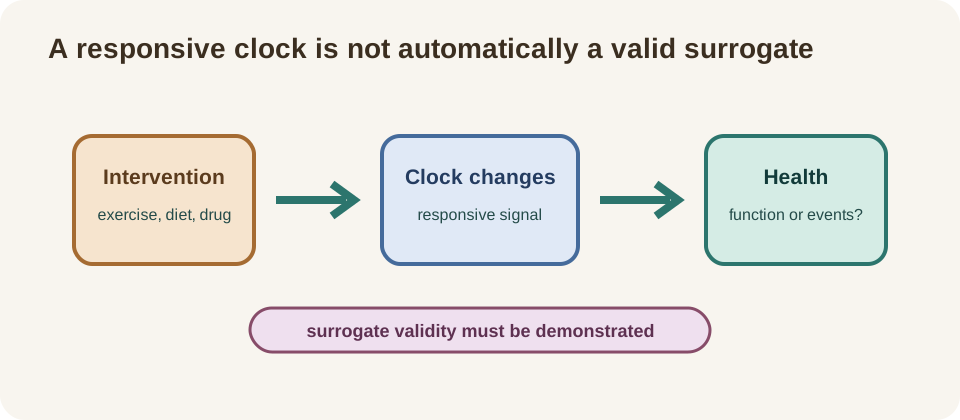
An August 2026 Nature Medicine analysis assembled the TranslAGE database of 51 longitudinal intervention studies and calculated a consistent panel of 16 prominent epigenetic clocks plus 94 other DNA-methylation biomarkers. The analysis reported that mortality- and pace-of-aging-oriented clocks showed stronger responses overall, while pharmacological and lifestyle interventions produced notable biomarker responses. Study population and duration also influenced responsiveness.
This is useful methods progress because fragmented intervention studies are difficult to compare when they use different clocks and assays. A harmonized analysis can identify patterns worth testing in prospective trials. It does not qualify any clock as a surrogate endpoint. A responsive biomarker must also reliably predict a meaningful patient outcome when an intervention changes it.
What this changes for biological-age research
The conceptual change is from asking for the one true biological age toward defining a measurement target and a biological context. Brain, immune, vascular and kidney signals may not move together. A multi-organ map could eventually support earlier detection or more tailored research, but organ age is not yet a routine clinical diagnostic.
The next phase needs longitudinal cohorts, repeated samples, harmonized assays, transparent model definitions and intervention studies with clinical or functional outcomes. Researchers should test whether a clock predicts outcomes better than established measures and whether using it changes care. They should also report null results, adverse signals, missing data and performance across populations.
- identify the target tissue, cell type and outcome;
- separate risk prediction from causal aging measurement;
- check repeatability, calibration and external validation;
- treat intervention responsiveness as a trial-design clue;
- do not call organ age a diagnosis or clock change a healthspan result.