What a proteomic aging clock measures
A proteomic aging clock uses patterns of circulating proteins and a statistical model to estimate an age-related score. Proteins sit closer to many downstream physiological processes than DNA methylation does, so they can reflect metabolism, inflammation, tissue injury, immune activity and signaling between organs. That proximity is useful, but it also makes the score sensitive to disease, medication, acute illness and technical platform effects. A 2026 analysis embedded six clocks in a randomized phase 2a trial, showing how they can be studied alongside treatment while leaving surrogate validity unresolved.
A clock estimate is not a blood-measured “true age.” It is a model output trained for a defined purpose in a defined cohort. The proteomic-aging-clocks canonical page keeps assay, model and population context attached to the number.
How a protein clock differs from a DNA-methylation clock
Both approaches use a model to compress many molecular measurements into a score, but they read different layers of biology. DNA-methylation clocks use chemical marks on DNA. Proteomic clocks use measured protein abundance, which can sit closer to active signaling, tissue injury, immune activity and organ function. That proximity may help a score reflect current physiology, while also making it sensitive to infection, treatment, kidney clearance and assay conditions.
A protein score and a methylation score can disagree without either being a direct measure of “true age.” They may capture different processes, time scales or technical features. The useful comparison is whether each model was trained and tested for the question being asked, not which one produces the most compelling number. The biological-age test guide explains why different test families should not be treated as interchangeable diagnoses.
The distinction also matters for intervention studies. Protein levels can change quickly with acute illness, medication or organ stress. A responsive assay may detect such change, yet researchers still need to show that it reflects a durable aging process and predicts an outcome people care about.
Why organ-specific clocks may add information
Chronological age and a single organism-level score can conceal heterogeneity. Two people with the same estimated biological age may have different brain, kidney, artery, muscle, liver or immune profiles. Organ-specific clocks attempt to separate those signals by using proteins enriched in particular tissues or systems.
The distinction is not absolute. Circulating proteins can be produced in several tissues, cleared by the kidney or liver, and altered by systemic inflammation. “Brain age” from plasma therefore does not mean a direct scan of neuronal age. It means that a protein pattern associated with brain-related biology predicts an age-related construct in the study’s model.
What the 2025–2026 Nature Aging work found
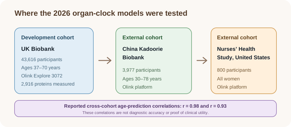
The large organ-specific study used plasma proteomics and machine learning to develop an organismal clock and ten organ-specific clocks in 43,616 UK Biobank participants. It used 2,916 measured proteins from the Olink Explore 3072 panel and identified 418 proteins enriched in at least one of ten organ or system categories.
The models were externally tested in 3,977 Chinese participants from the China Kadoorie Biobank and 800 U.S. women from the Nurses’ Health Study. The paper reported high cross-cohort performance, with correlations of 0.98 and 0.93 in the reported validation comparisons. The cohorts were not interchangeable: age range, sex composition, geography, ancestry, recruitment and platform context affect generalizability.
An independent East Asian study of proteomic organ-specific signatures provides an important replication direction. Replication across populations is valuable, but external prediction still answers a risk-stratification question rather than whether an intervention slows aging.
What the cross-cohort tests establish
The 2026 Nature Aging study developed its models in 43,616 UK Biobank participants, then tested them in 3,977 people in the China Kadoorie Biobank and 800 women in the U.S. Nurses’ Health Study. The participants differed in geography, age range and sex composition. All three cohorts used the Olink Explore 3072 panel, which measured 2,916 proteins. The investigators used tissue-expression data from GTEx to identify 418 proteins enriched in one or more of ten organ or system categories.
The reported cross-cohort correlations of 0.98 and 0.93 describe age-prediction performance across the tested cohorts. They do not mean the clocks classify disease with 98% or 93% accuracy. The study separately examined future disease and mortality associations, which are different validation questions. A model can estimate chronological age well and still have limited value for clinical decisions.
Organ age gaps were only weakly correlated with one another across cohorts. That pattern is consistent with the idea that a whole-body average may conceal differences between organ systems. It does not prove that each score measures aging intrinsic to that organ: circulating proteins may come from multiple tissues, and illness can alter the same signals. The organ-specific biological-age article discusses this distinction.
The authors reported that 121 of 187 organ–disease associations in the UK Biobank remained significant after false-discovery-rate correction, with many associations replicated in the China and U.S. cohorts. They also tested reduced protein panels, a step that may matter for future translation. A smaller panel could be easier to deploy, but feasibility does not replace prospective evidence that using a test improves care.
| Finding | What it supports | What it does not prove |
|---|---|---|
| High age-prediction correlation | the model tracks age in the tested cohorts | the score is a biological clock of causal aging |
| Organ age gap predicts disease | prospective risk association | the clock causes disease or diagnoses it |
| External validation | portability under tested conditions | universal performance in every population |
| Clock movement after treatment | possible intervention responsiveness | rejuvenation or longer life |
How the organ-clock evidence developed
A 2023 Nature study established an earlier multi-cohort foundation for organ-specific plasma-protein signatures. It analyzed 4,979 proteins in 5,676 adults across five cohorts, built models for eleven organs and reported that about one in five participants showed strongly accelerated aging in one organ, while about 1.7% met its multi-organ pattern. The study linked these profiles to disease and mortality associations.
The 2025–2026 work extends that line of research with larger datasets, additional external validation and newer organ-specific or cell-type-specific models. The result is a broader map of how protein patterns relate to systems and risk. This sequence shows methodological development; it is not evidence that measuring a clock and acting on it improves health.
Brain-age acceleration and mortality
In the Nature Aging study, brain-age acceleration showed the strongest association with all-cause mortality. The reported hazard ratio was 1.44 per standard-deviation increase in brain age gap in the discovery analysis. Brain age also related to neurodegenerative outcomes, while kidney, artery and heart signals aligned with their corresponding disease patterns.
These associations are biologically interesting because brain aging may connect vascular, immune and cognitive pathways. They remain associations, even when adjusted for clinical and lifestyle variables. A proteomic brain-age score should not be used to diagnose dementia, predict an individual’s exact lifespan or replace clinical assessment.
The organ-specific biological-age page explains why one organ score can be informative without being a literal tissue age.
Why the brain signal attracted attention
In the 2026 Nature Aging analysis, a one-standard-deviation higher brain age gap was associated with a 1.44-fold relative hazard of all-cause mortality in the UK Biobank analysis. The brain score was also associated with later neurodegenerative outcomes. These are group-level risk associations from observational follow-up, even though the models adjusted for several clinical, demographic and lifestyle factors.
Such results can help researchers prioritize pathways and design cohorts, but they do not supply a personal life-expectancy estimate. A brain-associated plasma pattern is not a neurological examination or imaging result. It cannot diagnose dementia, establish that a protein change caused disease, or show that lowering the score would reduce risk. Replication strengthens the evidence for prediction while leaving causal questions open.
Cell-type signatures, longitudinal change and platform effects
Newer work is moving from organ-level models toward cell-type-specific blood aging signatures. This may improve biological interpretability by connecting protein patterns with immune or other cellular states and with disease resilience. It also increases model complexity and the need for careful replication.
Repeated measurements can ask whether a person’s score changes over time, but cross-sectional age prediction is not the same as measuring an aging rate. A within-person change may reflect illness, treatment, weight, inflammation, hydration, batch effects or regression to the mean. A 20-year Nature Health cohort followed 1,298 people across four time points and measured 10,776 proteins, finding that proteins differed substantially in long-term stability. A panel of 21 proteins defined personalized baselines; deviations were associated with mortality in validation analyses. This supports studying stable personal profiles alongside age-prediction clocks, but it does not validate an intervention endpoint. The 2026 longitudinal organ-clock analysis in the brief’s source set remains preprint evidence and should be labeled preliminary.
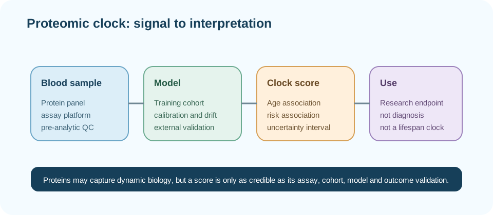
Platform matters. Aptamer-, antibody- and mass-spectrometry-based assays do not measure exactly the same protein universe or with identical error profiles. Calibration, age-bias correction, missingness and batch handling must be reported before results can travel between laboratories.
Cell-type clocks add a finer level of detail
A June 2026 Nature Medicine study analyzed more than 7,000 plasma proteins in 60,542 people and built protein-based aging models for more than 40 cell types. Its central idea is that blood proteins can carry signatures associated with particular cells, allowing researchers to ask whether aging-related patterns differ among immune, neuronal, glial, epithelial and other cell populations.
The authors reported that roughly 20–25% of people showed an accelerated pattern in one cell type, while 1–3% showed that pattern in ten or more cell types. These are model-defined categories, not cell-by-cell biopsies. The study linked selected cell-type scores to disease and mortality over follow-up, including an association between extreme astrocyte aging and Alzheimer’s disease risk among APOE4 homozygotes.
Cell-specific scores may help describe why people with similar chronological ages have different disease risks. The findings remain observational and depend on the cohort, assay platform, protein annotation and model thresholds. They generate testable hypotheses about vulnerability and resilience, but do not establish that a particular cell type causes an individual’s illness.
Prediction is not causal biology
A protein signature can predict future disease because it reflects early disease, a shared cause, treatment exposure or a consequence of another process. Statistical adjustment can reduce confounding but does not turn a prediction model into a causal mechanism. Genetic instruments, experimental perturbation and randomized intervention studies may help answer causal questions.
Organ-specific clocks may also be more predictive because they encode known clinical biomarkers or organ injury. That can be useful, but a powerful predictor is not necessarily a modifiable aging target. The clinical value depends on whether measuring it changes decisions and improves outcomes.
East Asian data broaden the validation picture
A 2026 EBioMedicine study used the prospective China Kadoorie Biobank to test organ-specific protein-age models in an East Asian population. It measured Olink and SomaScan proteins in about 4,000 participants with a mean age of 58 and derived age gaps for eighteen organs. The study examined associations with age-related traits and incident disease after accounting for multiple testing.
Its kidney score was associated with incident stroke, chronic liver disease and chronic kidney disease. The reported hazard ratios per one-year higher kidney ProtAgeGap were 1.03 for stroke, 1.11 for chronic liver disease and 1.19 for chronic kidney disease. The authors said these biomarker associations need further validation before clinical-trial use.
This is useful non-European evidence, but it is a separate analysis with its own model construction, platforms and outcomes. It supports the broad proposition that protein-age patterns can carry prospective risk information in another population. It does not prove that the UK Biobank model transfers unchanged or that the score is a causal target. The proteomic-aging-clocks overview can compare these study designs without treating them as one universal test.
Can proteomic clocks serve as intervention endpoints?
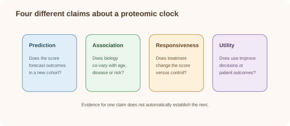
They could become useful endpoints if they are technically reliable, responsive to a prespecified intervention, reproducible across studies and demonstrably linked to a patient-important outcome. That last step is surrogate validation: the intervention’s effect on the clock must capture its effect on disease, function, quality of life or survival.
Evidence from caloric restriction, supplements or other interventions may show that a biomarker moves; it does not establish that the movement mediates healthspan. A September 2026 Nature Biotechnology analysis illustrates both promise and limits: six proteomic clocks were applied to serial serum samples from a randomized, double-blind, placebo-controlled phase 2a rentosertib trial in idiopathic pulmonary fibrosis. The parent trial enrolled 71 people, but the clock analysis included 42 with samples at all four time points (11 placebo and 31 across three treatment arms) over 12 weeks. Treated arms generally shifted toward lower predicted ages; 21 of 54 treatment-versus-placebo comparisons met the authors’ false-discovery threshold, with the clearest cross-clock signal at week 4. This small exploratory subset cannot separate aging biology from antifibrotic or disease-related changes and does not validate a clock as a surrogate. The TranslAGE brief makes the same distinction for DNA-methylation clocks: responsiveness is necessary, not sufficient.
For now, proteomic clocks are best treated as research measures that can refine hypotheses and trial design. They should not be marketed as clinical diagnoses or consumer instructions.
Repeated measurements still need outcome validation
Repeated sampling can show whether a score changes within a person, but it does not automatically distinguish durable aging change from acute illness, treatment exposure, natural fluctuation or measurement noise. A longitudinal organ-clock analysis listed in the brief is available as a preprint record; it should remain clearly labeled preliminary until peer review and independent replication.
For an intervention trial, investigators would need a prespecified clock, assay and analysis plan, repeated measurements, an appropriate control group and clinical outcomes measured over sufficient time. A favorable score shift is evidence of biomarker movement. It becomes a useful surrogate only if changes in that measure reliably track the intervention’s effects on disease, function, quality of life or survival across studies.
What consumer testing can and cannot tell you
A commercial proteomic report may provide a score, reference range or organ label, but the useful questions are which proteins were measured, on which platform, against which training cohort, with what calibration and uncertainty. A result can be technically impressive and still lack a validated action.
One number cannot summarize every organ, and a high or low score should not be interpreted without medical history, symptoms, medications and standard clinical testing. The biological-age-test page explains why these reports are not diagnoses.
Calibration determines whether a number can travel
Before comparing two proteomic-age reports, readers need to know whether they use the same assay platform, protein panel, preprocessing, reference population and age-bias correction. A score trained on one platform may not preserve its calibration on another. Even a standardized age gap depends on the model’s training data and the statistical method used to remove chronological age.
Those details affect scientific replication and consumer interpretation. A result outside a reference range may reflect a physiological signal, but it may also reflect platform differences, a population mismatch or a model that has not been validated for that use. A clinically useful test would need transparent uncertainty, reproducible measurements and evidence that reporting the score improves a decision or outcome. The epigenetic-aging overview offers a parallel explanation of how biomarker prediction differs from endpoint validation.
Evidence verdict
Proteomic aging clocks are an important 2026 research direction. Organ-specific models trained in large cohorts and tested in China and the United States show that circulating protein patterns can predict disease and mortality beyond chronological age. Brain-age acceleration stood out for mortality and neurodegenerative risk, while multiple organs contributed to broader disease patterns.
The conclusion remains bounded. Proteomic age is a model estimate, not a literal organ age; prospective prediction is not causation; and external validation is not universal clinical qualification. A 2026 phase 2a analysis shows that clocks can be added to a treatment trial, while a long-term proteome cohort offers a way to examine personal baselines. Neither establishes a validated surrogate. The field still needs assay harmonization, calibration, independent replication, longitudinal evidence and intervention studies tied to function, disease and survival.
The responsible conclusion is use proteomic clocks to improve research questions, not to diagnose aging or promise rejuvenation. The epigenetic-aging page and longevity clinical-trials page provide complementary endpoint context.
- name the assay, platform and cohort;
- separate age prediction from aging-rate measurement;
- report organ-specific signals with uncertainty and generalizability;
- treat clock movement as biomarker evidence, not lifespan proof;
- require clinical linkage before surrogate claims.